


AV and Semilunar Valves: A Cardiac Guide
.png)
At 7:30 a.m., a patient sits in front of you while your stethoscope warms in your hand. You hear a murmur, notice mild shortness of breath, and need to decide whether the sound reflects a normal flow pattern, a narrowed valve, or blood moving backward. That bedside judgment starts with understanding the AV and semilunar valves.
This guide builds the subject from structure to sound, then from sound to pathology and modern imaging decisions. You'll learn where each valve sits, which pressure phase opens or closes it, how stenosis and regurgitation present, and why clinicians sometimes need more than a routine echocardiogram.
Why These Four Valves Matter at the Bedside
The heart has four valves, divided into two functional groups. The mitral and tricuspid valves are the atrioventricular, or AV, valves. They sit between the atria and ventricles. The aortic and pulmonary valves are the semilunar valves, positioned between the ventricles and the great arteries. This four-valve arrangement maintains one-way blood flow through the heart. A detailed review of valve structure and function describes how these valves open and close in response to changing pressure.
Think of each valve as a pressure-controlled door. It doesn't open because a muscle actively pulls it open. It opens when pressure behind it exceeds pressure ahead of it. It closes when the pressure relationship reverses.
That simple rule explains much of what you hear and see:
- AV valves open during ventricular filling, allowing blood to move from the atria into the ventricles.
- AV valves close during ventricular contraction, preventing blood from returning to the atria.
- Semilunar valves open during ventricular ejection, allowing blood into the aorta and pulmonary artery.
- Semilunar valves close during relaxation, preventing blood from returning to the ventricles.
At the bedside, accuracy depends on connecting three questions:
- Where is the valve?
- What pressure phase controls its movement?
- What happens when the valve becomes narrow or leaky?
A narrowed valve creates resistance. A leaky valve permits backward flow. Both problems can alter chamber pressures, change blood flow, produce a murmur, and eventually affect symptoms or organ perfusion.
Practical rule: Don't memorize a murmur in isolation. First identify the valve, then determine whether blood is struggling to move forward or escaping backward.
The rest of the examination follows that logic. Anatomy tells you what the valve is built to do. The cardiac cycle explains when it moves. Auscultation reveals the mechanical consequence, while echocardiography and advanced imaging help determine whether the finding is clinically important.
Anatomy of the AV and Semilunar Valves
A patient's murmur becomes easier to localize once you can picture the four valves in relation to the chambers and great vessels.
- Mitral valve: Separates the left atrium from the left ventricle.
- Tricuspid valve: Separates the right atrium from the right ventricle.
- Aortic valve: Directs blood from the left ventricle into the aorta.
- Pulmonary valve: Directs blood from the right ventricle into the pulmonary artery.
The AV valves include more than their visible leaflets. Each leaflet connects through chordae tendineae to papillary muscles. Together, these structures resemble a parachute's cords and anchors. As the ventricles contract, papillary muscles tighten the chordae and help keep the leaflets from turning inside out into the atria. They support closure rather than actively opening the valves.
The mitral valve has two main leaflets, whereas the tricuspid valve has three. The semilunar valves are built from three crescent-shaped cusps and lack chordae tendineae and papillary muscles. Their cusps meet at commissures, creating a seal when pressure in the arteries exceeds ventricular pressure. Normal valve tissue is typically less than 1 mm thick. AV valves are slightly thicker than semilunar valves, and left-sided valves are slightly thicker than right-sided valves. This structural comparison describes these differences.
Finding the valves on the chest
Auscultation sites are listening windows, not the valves' exact surface locations. Sound travels with blood flow and is often clearest downstream from the anatomical valve.
- Aortic area: Right second intercostal space near the sternal border.
- Pulmonic area: Left second intercostal space.
- Tricuspid area: Lower left sternal border.
- Mitral area: Cardiac apex, usually near the left fifth intercostal space.
The fibrous skeleton forms a firm framework around the valve openings and electrically separates atrial from ventricular muscle. The aortic cusps also lie near the coronary ostia. Aortic-root disease or an intervention there can therefore affect coronary blood supply as well as valve function.
For learners linking valve anatomy with emergency cardiovascular assessment, ACLS Certification is available online through ProMed Certifications. Course information states that learners can study at a convenient time and place, complete the exam, and receive their card in as quick as one hour or less.
How the Valves Work in the Cardiac Cycle
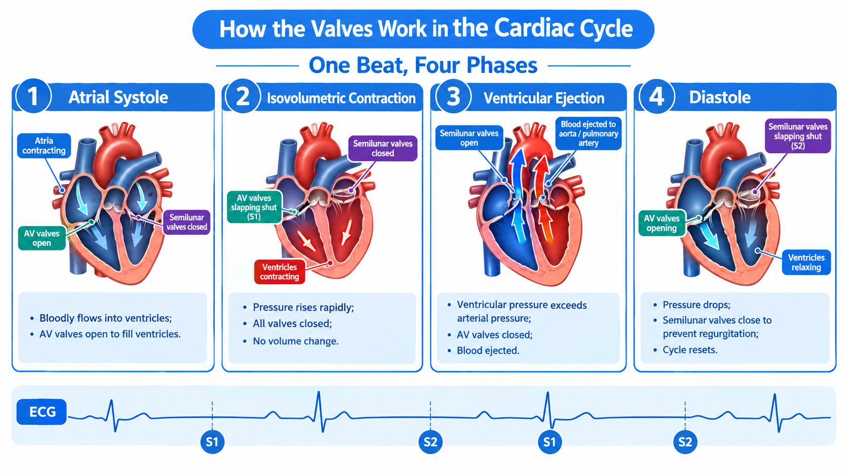
Follow one heartbeat as a sequence of pressure changes rather than as four disconnected facts.
During atrial systole, the atria contract and push blood into the ventricles. The mitral and tricuspid valves are open. The aortic and pulmonary valves remain closed because ventricular pressure hasn't yet exceeded arterial pressure.
The electrical signal then travels through the ventricles. Around the QRS complex, ventricular depolarization triggers contraction. Ventricular pressure rises sharply. Once it exceeds atrial pressure, the AV valves close. This produces S1, the first heart sound. For a brief interval, all four valves are closed. The ventricles are contracting, but no blood enters or leaves, so this is called isovolumetric contraction.
When ventricular pressure becomes greater than pressure in the aorta and pulmonary artery, the semilunar valves open. Blood is ejected. The AV valves stay closed, protecting the atria from ventricular pressure.
As ventricular repolarization progresses around the T wave, contraction weakens. Aortic and pulmonary artery pressure becomes higher than ventricular pressure, forcing the semilunar valves shut. Their closure produces S2. Isovolumetric relaxation follows, with all valves closed again.
When ventricular pressure falls below atrial pressure, the AV valves reopen. Rapid ventricular filling begins, followed by slower filling. The cycle then returns to atrial systole.
The electrical and mechanical loop
The ECG shows electrical timing. The pressure curves show mechanical response. Valve sounds mark the points where pressure closes the doors.
For a practical refresher on linking ECG timing to cardiac mechanics, use this guide to reading ECGs. The key is to listen while asking what the ventricles are doing at that instant, not just to label a sound as “lub” or “dub.”
What Each Heart Sound Tells You
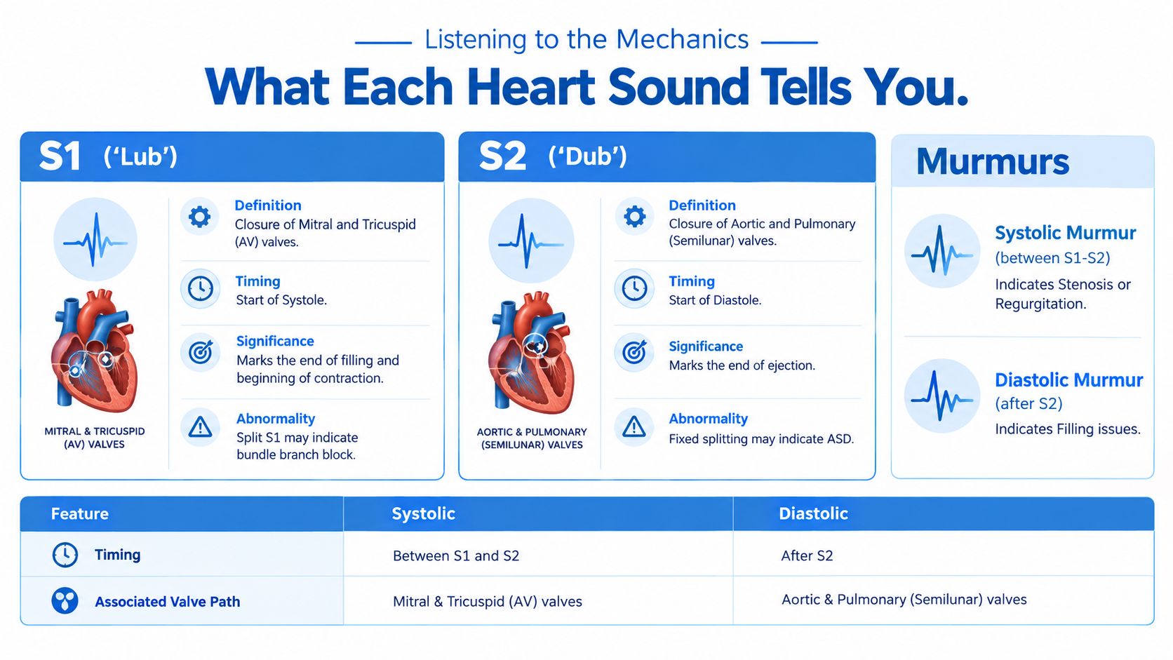
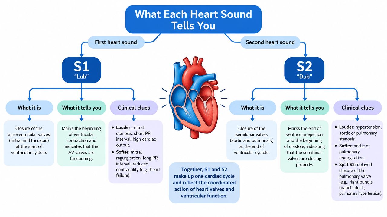
S1 is the sound of the mitral and tricuspid valves closing. It marks the beginning of ventricular systole. S2 is the sound of the aortic and pulmonary valves closing, marking the beginning of diastole.
You may hear physiologic splitting of S2 during inspiration. The pulmonic component, P2, can occur after the aortic component, A2, because inspiration changes right-sided filling and prolongs right ventricular ejection. A persistently fixed split has a different clinical meaning and should prompt consideration of conditions such as an atrial septal defect.
Match the sound to the listening point
Use a deliberate sequence during auscultation:
- Aortic area: Listen for ejection sounds and murmurs caused by obstruction across the aortic valve.
- Pulmonic area: Assess P2, splitting of S2, and right-sided outflow murmurs.
- Tricuspid area: Consider tricuspid regurgitation, especially when right-sided volume or pressure is increased.
- Mitral area: Listen for mitral regurgitation, mitral stenosis, S3, and S4.
A murmur between S1 and S2 is systolic. It may reflect stenosis of an outflow valve or regurgitation through an AV valve. A murmur after S2 is diastolic. It may reflect regurgitation through a semilunar valve or restricted filling through an AV valve.
S3 occurs during rapid ventricular filling. It can be physiologic in younger patients, but in an older adult it may raise concern for impaired ventricular function and heart failure. S4 occurs when the atria contract against a stiff ventricle, often described as an “atrial kick” sound.
A patient with pericardial fluid may present with findings that overlap with other causes of reduced cardiac filling. Review cardiac tamponade assessment when the clinical picture includes hypotension, high venous pressure, or muffled heart sounds.
For clinicians completing ongoing education, ProMed+ Physician CME provides on-demand clinical curricula and access to more than 200 AMA PRA Category 1 Credits™ according to the catalog information.
Stenosis vs Regurgitation Across the Four Valves
Stenosis means the valve opening is restricted. The chamber behind the valve must generate more pressure to move blood forward. Regurgitation means the valve fails to close completely, allowing blood to move backward.
Aortic stenosis produces a classic crescendo-decrescendo systolic ejection murmur. As the valve narrows, left ventricular ejection becomes more difficult. Syncope, angina, and dyspnea are the classic symptom pattern, and symptomatic severe disease generally requires prompt evaluation for valve replacement.
Mitral stenosis produces an opening snap followed by a diastolic rumble. Rheumatic disease remains a strong historical association. Mitral regurgitation is usually holosystolic and may be primary, caused by leaflet or chordal disease, or secondary, caused by ventricular or annular changes.
Aortic regurgitation is diastolic because blood returns from the aorta into the left ventricle after the aortic valve should have closed. Acute regurgitation can cause rapid hemodynamic deterioration. Chronic regurgitation allows adaptation for a time, but progressive ventricular enlargement or symptoms change the urgency.
Tricuspid regurgitation is easy to overlook. Look for raised jugular venous pressure, hepatic congestion, and other signs of right-sided volume overload. Pulmonic valve disease is uncommon outside congenital disease or severe pulmonary hypertension.
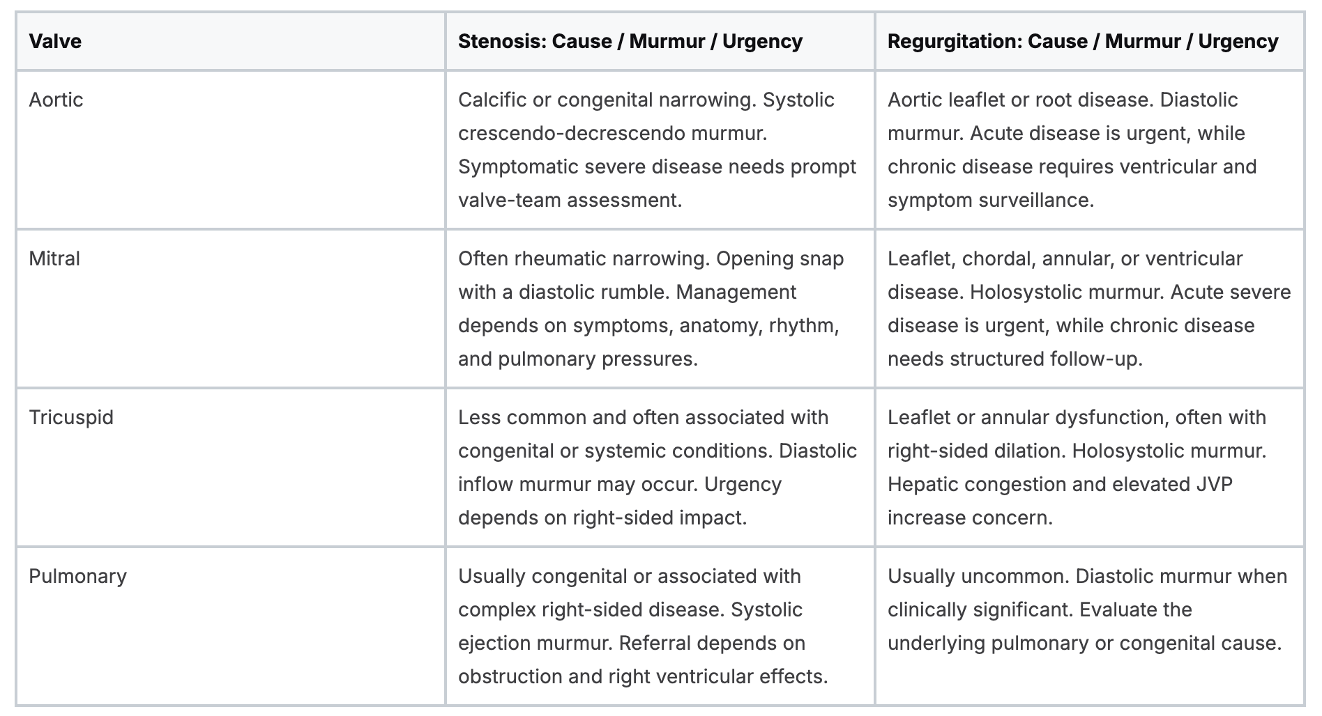
The same sound can therefore represent different lesions depending on timing, location, radiation, and the patient's symptoms.
From Echo to Decisions on Subtle Valve Findings
Transthoracic echocardiography is the usual first-line test for valve assessment. It shows leaflet motion, chamber size, ventricular function, flow velocity, pressure gradients, and regurgitation. The difficult cases arise when these measurements disagree with one another or don't fit the patient's symptoms.
A borderline aortic stenosis study may require more than one number. Clinicians consider valve area, flow state, ventricular ejection fraction, gradients, and leaflet appearance. Low-flow, low-gradient aortic stenosis can occur with preserved or reduced ejection fraction, and the distinction changes the next diagnostic step.
When echocardiography doesn't settle the question
- Transesophageal echocardiography: Useful when mitral anatomy, prosthetic valves, or leaflet detail requires closer visualization.
- Stress echocardiography: Helps evaluate selected low-gradient aortic stenosis cases by observing valve and ventricular behavior under increased demand.
- Cardiac CT: Helps quantify aortic valve calcium when stenosis severity remains discordant.
- Cardiac MRI: Helps measure regurgitant volumes when echocardiographic estimates remain uncertain.
Recent ESC/EACTS guidance highlights sex-specific CT calcium thresholds for aortic stenosis. A threshold above 1200 AU in women and above 2000 AU in men supports severe aortic stenosis in the appropriate clinical context. The guideline discussion emphasizes that imaging should be interpreted as an integrated decision, not as an isolated measurement.
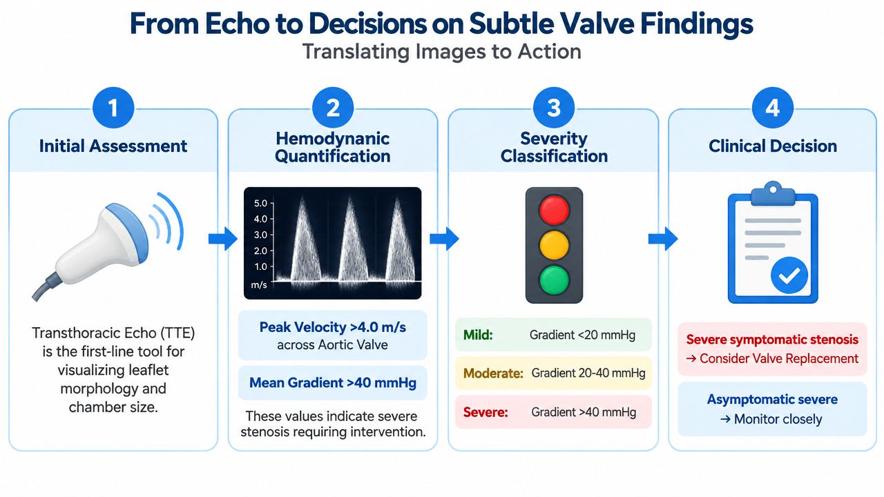
Patients with mild, borderline, or asymptomatic disease often need serial surveillance, not immediate intervention. The 2025 ESC/EACTS framework stresses integrative assessment and continued follow-up rather than relying on a single metric. This review of surveillance and screening questions also notes that broad screening of asymptomatic older adults remains controversial.
A careful home history can support, but never replace, clinical evaluation. Patients may find it useful to monitor cardiac health yourself by tracking symptoms and functional changes to discuss with a clinician.
Treatment Options and Key Historical Milestones
Valve treatment is a continuum. An asymptomatic patient with mild disease may need observation and repeat imaging rather than a procedure. When symptoms, ventricular changes, severe obstruction, or progressive regurgitation appear, the care team may consider medical therapy, repair, replacement, or a transcatheter approach.
Medical therapy treats the consequences and associated conditions. Rate control can help patients with mitral stenosis and atrial fibrillation. Afterload reduction may support selected patients with regurgitation. These treatments don't repair a severely damaged valve, so clinicians still monitor anatomy, ventricular response, symptoms, and rhythm.
Repair preserves native tissue when anatomy permits. Replacement may use a mechanical prosthesis or a bioprosthesis. Mechanical valves offer durability but require long-term anticoagulation. Bioprosthetic valves may reduce anticoagulation demands but have different durability considerations. The choice depends on anatomy, age, surgical risk, pregnancy considerations, anticoagulation tolerance, lifetime planning, and patient preference.
Historical milestones show how quickly treatment has evolved. The Hufnagel caged-ball valve appeared in 1952, and Starr and Edwards introduced early ball-in-cage prostheses, replacing the first mitral valve in 1961 and the first aortic valve in 1962. The Ross procedure, described in 1967, uses the patient's pulmonary valve to replace a diseased aortic valve. The first TAVR was performed in 2002, followed by formal U.S. FDA approval in 2010.
Balloon valvuloplasty remains useful in selected rheumatic and pulmonic lesions. Transcatheter options now include TAVR for aortic stenosis and edge-to-edge repair approaches for selected mitral and tricuspid regurgitation. To understand the wider clinical team behind these interventions, review how to become a cardiovascular perfusionist.
Keeping Your Valve Knowledge Sharp and Where to Go Next
Competence with valve disease doesn't require a cardiology fellowship or an in-person American Heart Association class. Online CE and certification programs are valid, accessible, and can be just as effective as in-person education when the curriculum, assessment, and accreditation are sound. The belief that only AHA or American Red Cross in-person classes count is outdated. Acceptance still depends on employer and licensing requirements, so verify those requirements before enrolling.
The evidence supports a balanced view. A systematic review of online learning in health professions found changes in knowledge, skills, attitudes, and satisfaction, with high- and low-quality studies indicating equivalence and possible advantages over traditional formats for knowledge and skills gained. This review supports online CE as a credible route for flexible professional development.
A Cochrane Review reached a more cautious conclusion, finding that e-learning may make little or no difference compared with traditional learning for patient outcomes, professional behaviors, and knowledge. Its findings reinforce the useful point that neither format is automatically superior. Course design matters.
Spaced education is one example of that design principle. A 2024 meta-analysis found spaced online education superior to massed online education for post-intervention knowledge, with a standardized mean difference of 0.32 and a 95% confidence interval of 0.13 to 0.51. The meta-analysis also reported improvements in knowledge, skills, confidence, and clinical practice change.
Build your own learning loop:
- Choose one valve: Review one anatomy diagram and a short echo clip each week.
- Explain it clearly: Write a two-sentence summary in your own words.
- Test recall: Identify the valve, timing, sound, and likely lesion during rounds.
- Use dependable references: Keep the ASE guidelines, British Society of Echocardiography modules, Mayo Clinic Proceedings cases, NEJM Images in Clinical Medicine, UWorld, and TrueLearn within your study system.
- Follow one patient longitudinally: Compare symptoms, examination findings, and imaging over time.
Online CE and certifications are gaining acceptance across healthcare institutions because clinicians need learning that fits real schedules without sacrificing quality. A 2024 scoping review found improvements in knowledge, skills, performance, confidence, self-efficacy, satisfaction, and attitudes across online and blended formats. The review supports the broader move toward flexible continuing professional development.
ProMed Certifications offers online medical certification courses, including ACLS, BLS, PALS, CPR, and neonatal resuscitation, with self-paced access and certification pathways described on its platform. Visit ProMed Certifications to review the available courses, select the training that matches your role, and continue building practical cardiovascular knowledge around your schedule.
.avif)
Stay compliant with ProMed+
Certifications included: ACLS, BLS, PALS, CPR & Neonatal Resuscitation
Unlimited continuing education: over 200 hours of accredited CME
All-inclusive: One price. No surprises.
Get certified today

The Advanced Cardiac Life Support (ACLS) is an accredited online medical certification course that teaches medical professionals to respond to nearly all cardiopulmonary emergencies.

The Pediatric Advanced Life Support (PALS) Recertification instructs medical professionals on performing pediatric cardiopulmonary resuscitation in emergencies.

The Basic Life Support (BLS) Recertification is intended to teach healthcare professionals the basic steps of CPR and rescue breathing for adults, children, and infants.

Our online medical certification course for CPR, Automated External Defibrillator (AED), and First Aid is designed to teach adult, child, and infant CPR and AED use.

The Neonatal Resuscitation Certification trains medical professionals in performing resuscitation procedures and life-saving techniques for newborns at the time of delivery.

.png)
.png)
.png)