


Heart Block Mobitz 2: What You Need to Know
.png)
You're reviewing a telemetry strip when the monitor suddenly skips a ventricular beat. The alarm sounds, the patient looks toward you, and someone asks whether it's artifact. That moment matters. A dropped QRS can reflect a dangerous failure of cardiac conduction, especially when the preceding PR intervals remain unchanged.
Heart block Mobitz 2, also called Mobitz type II second-degree atrioventricular block, is an important rhythm for nurses, physicians, paramedics, and other healthcare professionals to recognize quickly. This guide builds from the basic ECG pattern to the harder cases, including 2:1 conduction, intermittent block, and broader conduction disease, then connects each finding to the question that changes management: does this patient need pacing now?
A Rhythm That Demands Your Attention
Start with the patient, not the monitor. When a telemetry alarm reports a pause or dropped beat, assess the pulse, blood pressure, mental status, skin temperature, and overall perfusion. A patient who is hypotensive, confused, pale, short of breath, or experiencing chest pain needs immediate escalation while another clinician confirms the rhythm.
A single nonconducted P wave in Mobitz II carries more concern than a typical dropped beat in Mobitz I. In Mobitz II, the atrial impulse appears to conduct normally, then suddenly fails below the AV node, usually in the His–Purkinje system. The next ventricular beat may depend on an escape rhythm that's slow and unreliable. Merck Manual's overview of atrioventricular block explains that progression can be sudden and that the ventricular escape rhythm may fall to 20 to 40 beats per minute.
The first bedside checks
Use a short, disciplined sequence:
- Confirm the patient: Check responsiveness, breathing, pulse, and the possibility of loose leads or signal artifact.
- Assess circulation: Measure blood pressure and look for signs of poor perfusion.
- Find the P wave: Determine whether an atrial impulse appears without a following QRS.
- Call for help early: Notify the responsible clinician and prepare for higher-acuity monitoring.
- Anticipate pacing: Keep emergency pacing equipment available when Mobitz II is suspected.
A stable blood pressure doesn't make the rhythm harmless. The block can be intermittent, so the patient may look well between pauses. The dangerous feature is the unpredictable loss of ventricular conduction, not merely the symptoms present during the strip.
Practical rule: Treat a suspected Mobitz II pattern as a warning sign until a qualified clinician confirms the rhythm and identifies the cause.
The 2018 ACC/AHA/HRS bradycardia guideline classifies permanent pacing as a Class I recommendation for acquired Mobitz II that isn't caused by a reversible or physiologic condition, even when symptoms are absent. That recommendation changes the next steps: continuous monitoring, cardiology involvement, and prompt pacing assessment are more appropriate than casual observation.
What Heart Block Mobitz 2 Actually Is
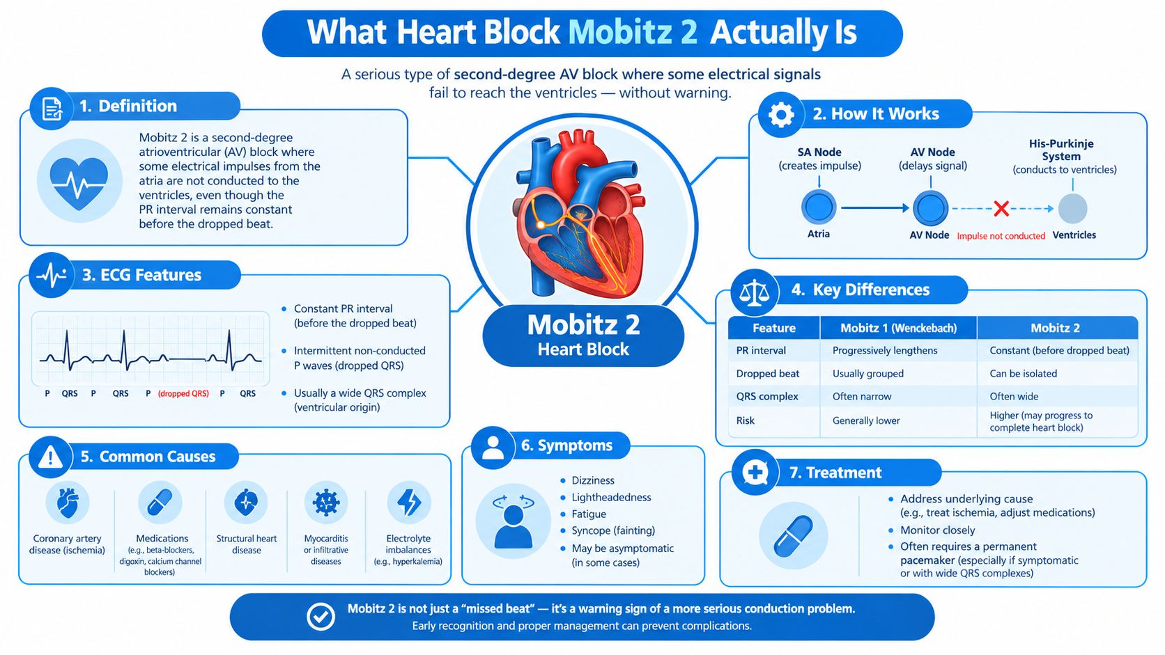
Second-degree AV block means some atrial impulses reach the ventricles while others don't. The atria continue producing P waves, but at least one P wave fails to generate a QRS complex.
Mobitz II has a specific visual rule. The PR interval stays constant on conducted beats, then a P wave suddenly appears without a following QRS. The atrial rhythm usually continues at a regular pace, so the P waves “march through” the pause.
Think of the conduction system as a relay route:
- The SA node starts the electrical signal.
- The atria carry that signal and produce the P wave.
- The AV node provides a controlled delay.
- The His bundle and bundle branches deliver the impulse toward the ventricles.
- The Purkinje network spreads activation through ventricular muscle.
In Mobitz II, the signal reaches the relay system, but a downstream station abruptly refuses to pass one impulse. There's no gradual PR lengthening before the failure. The conduction is all or nothing.
Why the QRS often looks wide
Mobitz II is usually an infranodal conduction disorder, meaning the problem lies below the AV node, often in the His–Purkinje system. The QRS is frequently widened because disease commonly involves the bundle branches. The American Heart Association and ACC guideline summary describes the defining pattern as a sudden nonconducted P wave with a constant PR interval, provided the sinus rate is stable and enough conducted beats are available for comparison.
Possible causes include degenerative conduction-system disease, anterior myocardial infarction, cardiomyopathy, infiltrative disease, medication toxicity, and injury after cardiac surgery or ablation. Reversible causes still matter. A rhythm that resembles Mobitz II can require a different plan if a medication, metabolic problem, ischemia, or physiologic mechanism is responsible.
Clinical teams also need operational support around cardiac services, from accurate documentation to reimbursement workflows. A resource on Happy Billing cardiology can help practices understand the administrative side of cardiology care. For clinicians building emergency-response knowledge, ACLS Certification is an online option that ProMed Certifications describes as a convenient way to complete an ACLS course, exam, and card process within a busy schedule.
Mobitz I vs Mobitz II at a Glance
The fastest way to separate Mobitz I from Mobitz II is to examine what happens to the PR interval before the dropped beat. In Wenckebach, the PR interval progressively lengthens until one P wave fails to conduct. In Mobitz II, the PR interval remains stable, then conduction stops unexpectedly.
The difference reflects anatomy. Mobitz I usually involves the AV node, where conduction can slow in a progressive, decremental fashion. Mobitz II generally reflects disease in the His–Purkinje system, where a damaged pathway can fail abruptly.
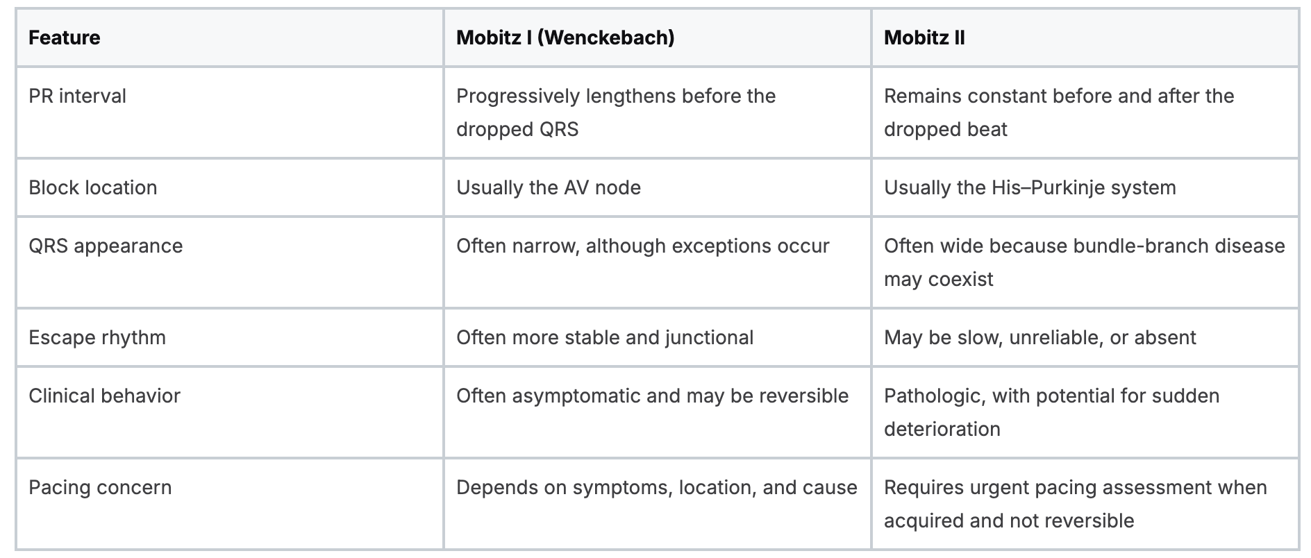
A narrow QRS favors nodal disease, while a wide QRS raises concern for infranodal disease. Neither feature should be used alone. A bundle-branch pattern may widen the QRS independently, and infranodal disease can occasionally present with a narrow QRS.
Why the distinction changes disposition
Mobitz I can remain stable, particularly when the patient is asymptomatic and the pattern fits a nodal mechanism. Mobitz II is different because the next blocked impulse may arrive without warning. The MedPrep review of second-degree heart block describes Mobitz II as a substantial subset of second-degree AV block, approximately 20% to 30%, and reports association with bundle branch block in approximately 70% to 80% of cases.
That same review reports a 35% to 50% risk of progression to complete heart block without pacing. Exact population prevalence remains difficult to establish because large population-based studies of Mobitz I and II are limited. The practical message is clearer than the epidemiology: a confirmed, acquired Mobitz II pattern shouldn't be managed like an incidental benign pause.
For healthcare professionals, online education supports this kind of pattern recognition without requiring travel or a classroom schedule. A systematic review of online eLearning in health professions education found changes in knowledge, skills, attitudes, and satisfaction, with evidence indicating that online learning can be equivalent to, and possibly more effective than, traditional learning for knowledge and skills in health professions education. That supports the practical value of accredited online CE and certification alongside in-person training.
Reading the ECG Strip With Confidence
Read the strip in a fixed order. First, identify the atrial rhythm. Then compare the PR intervals of conducted beats. Finally, search for a P wave that lacks a QRS, rather than assuming a pause represents sinus slowing.
A classic Mobitz II pattern
On a typical strip, the sequence looks like this:
- A P wave is followed by a QRS.
- The next PR interval is similar to the first.
- Another conducted beat follows with the same PR timing.
- A P wave appears, but no QRS follows.
- The next conducted beat resumes with the same PR relationship.
The blocked P wave may be hidden in the preceding T wave. Slow the paper or monitor display if possible, and inspect the T-wave contour carefully. The P-P intervals should remain regular. If the atrial timing pauses as well, consider sinus pause or sinoatrial dysfunction instead.
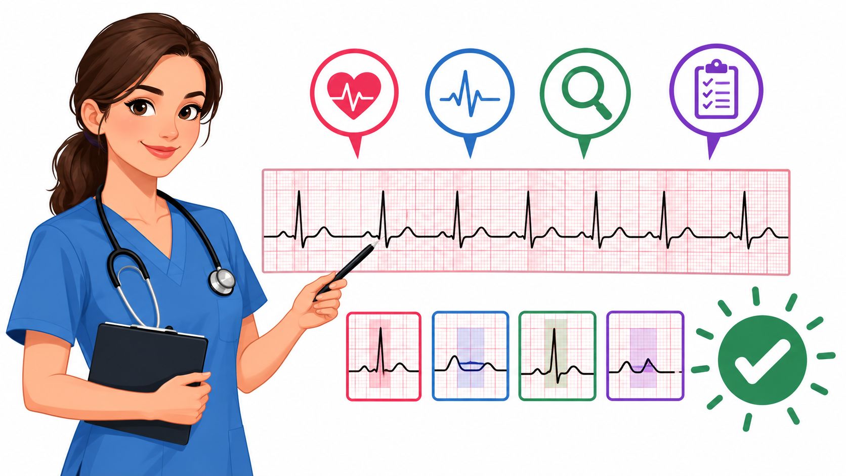
A structured introduction to interval measurement and waveform identification is available in this guide to reading ECGs. The key is repetition. You're training your eyes to track atrial activity independently from ventricular activity.
The difficult 2:1 pattern
In 2:1 AV block, every other P wave fails to conduct. That creates one conducted beat followed by one blocked atrial impulse. Because there aren't two conducted beats in a row, you can't reliably see progressive PR prolongation or prove PR stability from the surface pattern alone.
Use the QRS width, existing bundle-branch disease, clinical setting, and additional ECG evidence as clues. A wide QRS makes infranodal disease more concerning, but it doesn't establish the diagnosis by itself. Fixed ratios such as 3:2, 4:3, and 2:1 describe how many atrial impulses conduct, while high-grade block describes more extensive failure of conduction.
Common reading traps
- Buried P waves: Look inside T waves during the expected atrial cycle.
- Junctional escape: A rescue beat can appear without a preceding sinus P wave.
- Premature atrial contractions: An early P wave may be blocked and mimic a conduction pause.
- Lead artifact: Confirm the finding on another lead and, when possible, a full 12-lead ECG.
- Variable conduction: Don't force a rhythm into Mobitz I or II when the strip doesn't provide enough consecutive conducted beats.
For clinicians completing resuscitation education, BLS Certification is another online option from ProMed Certifications. Online CE and certifications are valid, accessible, and increasingly accepted when the course meets the relevant institutional, regulatory, and accreditation requirements. The format can save travel time and fit shift work without lowering the quality of the learning experience.
Clinical Presentation and How Patients Feel
Mobitz II can be silent or dramatic. Some patients have an abnormal ECG discovered during routine evaluation. Others describe dizziness, exercise intolerance, presyncope, or a sudden blackout that gives little warning.
The abruptness matters. In a gradual nodal pause, the patient may notice increasing lightheadedness. In Mobitz II, the conduction failure is sudden, so the ventricular rate can drop before the body compensates. A patient may lose consciousness briefly, fall, or sustain a Stokes–Adams attack.
Symptoms should be matched with the rhythm and the patient's circulation:
- Presyncope: A feeling that consciousness is about to disappear.
- Syncope: Sudden, temporary loss of consciousness from inadequate cerebral perfusion.
- Exercise intolerance: Reduced ability to increase cardiac output during activity.
- Chest discomfort or breathlessness: Possible signs of inadequate forward flow or underlying ischemia.
- No symptoms: Not reassuring when the ECG shows acquired Mobitz II.
Escape rhythm and stability
The escape rhythm provides a limited safety net below the block. A narrow escape generally originates higher in the conduction system and tends to be faster and more dependable. A wide escape usually reflects a more distal fascicular or ventricular origin, which can be slower and less reliable.
This differs from complete heart block. In complete block, atrial impulses continuously fail to conduct, and the escape rhythm becomes the ongoing ventricular rhythm. Mobitz II is intermittent, but that intermittent behavior can be more deceptive because the patient may appear stable before an unexpected pause.
The clinical team should also consider the information systems surrounding patient monitoring. When a hospital evaluates a healthtech engineering partner, reliable integration can support the flow of telemetry data, alerts, and documentation. Technology doesn't replace bedside assessment, but it can help clinicians receive and interpret important information without unnecessary delay.
For ongoing professional development, ProMed+ Nursing CE presents an online continuing-education platform for nurses, with streaming access to CE content and certificates described in its catalog. Online education works especially well for rhythm review because learners can revisit difficult ECG examples at their own pace.
Acute Care and Long Term Pacing Decisions
Management begins with stability. If the patient has hypotension, altered mental status, ischemic chest pain, acute heart failure, or other signs of poor perfusion, activate the local bradycardia response and prepare pacing while senior clinical support arrives.
Transcutaneous pacing may provide immediate support. Transvenous pacing can offer a more reliable bridge when the patient remains unstable or the rhythm is recurrent. Atropine may be ineffective because Mobitz II usually occurs below the AV node. Depending on local protocol and clinician judgment, isoproterenol or dopamine may serve as temporary bridges while definitive treatment is arranged.
Search for reversible causes
Pacing decisions shouldn't skip the diagnostic search. Review the clinical context and look for:
- Ischemia: Evaluate for acute coronary disease or infarction.
- Electrolyte disturbance: Check for hyperkalemia and other metabolic contributors.
- Medication toxicity: Review beta-blockers, calcium channel blockers, digoxin, and antiarrhythmic agents.
- Inflammatory or infectious disease: Consider Lyme carditis when the history supports it.
- Recent procedures: Ask about cardiac surgery or catheter ablation near the conduction system.
If a reversible cause explains the block, correcting that cause becomes central. If the block persists, or if the cause isn't reversible, the guideline-based pathway changes toward permanent pacing.
When permanent pacing becomes the plan
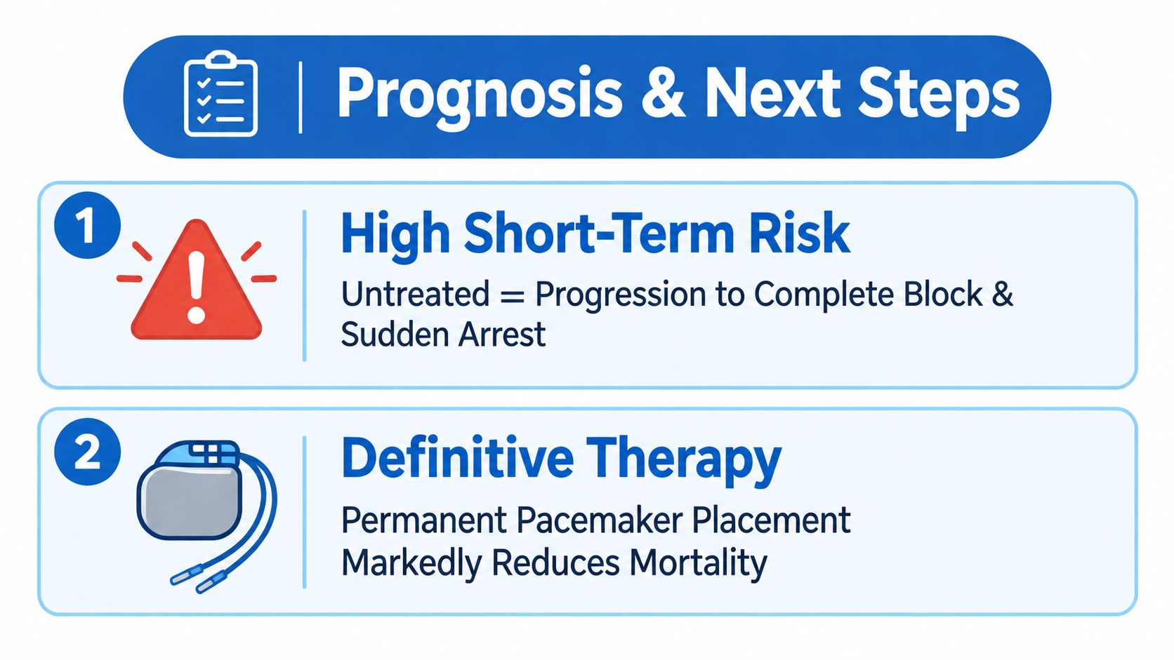
The 2018 ACC/AHA/HRS guideline recommends permanent pacing for acquired Mobitz II that isn't caused by a reversible or physiologic condition, regardless of symptoms. The guideline also treats high-grade AV block and complete AV block as serious pacing conditions. The 2021 ESC guidance similarly supports pacing for Mobitz II when symptoms or relevant risk factors are present, while newer appropriate-use discussions continue to treat advanced second-degree block as a pacing indication even without symptoms.
A monitored bed, cardiology consultation, and careful device planning are appropriate. Device selection, including single- versus dual-chamber pacing, depends on the patient's atrial rhythm, conduction pattern, comorbidities, and clinical context.
Clinicians who want a focused emergency reference can review the ACLS bradycardia with a pulse algorithm. For physicians managing licensure and clinical learning, ProMed+ Physician CME lists an on-demand library with unlimited access to more than 200 AMA PRA Category 1 Credits™, as described in its catalog. Online CME is now a credible complement to classroom training, and many healthcare organizations are recognizing its flexibility and equivalent educational value.
Prognosis and What You Should Do Next
Mobitz II is not a rhythm to watch casually. Without pacing, progression to complete heart block can be sudden, and the escape rhythm may fail to maintain adequate circulation. The 2024 review of Mobitz II emphasizes that the condition usually reflects His–Purkinje disease and commonly occurs with bundle branch block in approximately 70% to 80% of cases.
Use this practical sequence when the rhythm appears:
- Confirm the pattern: Obtain a full 12-lead ECG and compare conducted PR intervals.
- Assess the patient: Check symptoms, pulse, blood pressure, mental status, and perfusion.
- Find reversible causes: Investigate ischemia, hyperkalemia, medication effects, infection, and recent procedures.
- Arrange monitored care: Involve cardiology and prepare for pacing if the block persists or the cause isn't reversible.
The single ECG clue that should change your next decision is simple: a P wave fails to conduct despite constant PR intervals on the conducted beats. In 2:1 block, classification may remain uncertain, so the broader conduction pattern and clinical risk deserve particular attention.
Medication safety also matters during bradycardia assessment. Reviewing medications commonly found on a crash cart can reinforce what each emergency drug does, while local protocols and physician direction should guide actual treatment.
Online CE and certification are no longer limited to a classroom or to AHA and American Red Cross in-person classes. Accredited online programs can offer the same core educational purpose with greater flexibility, efficient review, and access for professionals working rotating shifts. Peer-reviewed reviews and meta-analyses support online learning across knowledge, skills, behavior, and learner outcomes, including evidence that spaced online learning can outperform massed online learning for post-intervention knowledge with an SMD of 0.32, with a 95% confidence interval of 0.13 to 0.51. The industry is gradually accepting online CE and certifications more broadly, and ProMed Certifications is one option for healthcare professionals who want future-focused, accessible education.
ProMed Certifications offers online ACLS, BLS, and continuing-education courses that let healthcare professionals study around demanding schedules. Visit ProMed Certifications to explore training that strengthens rhythm recognition, emergency readiness, and ongoing professional competence.
.avif)
Stay compliant with ProMed+
Certifications included: ACLS, BLS, PALS, CPR & Neonatal Resuscitation
Unlimited continuing education: over 200 hours of accredited CME
All-inclusive: One price. No surprises.
Get certified today

The Advanced Cardiac Life Support (ACLS) is an accredited online medical certification course that teaches medical professionals to respond to nearly all cardiopulmonary emergencies.

The Pediatric Advanced Life Support (PALS) Recertification instructs medical professionals on performing pediatric cardiopulmonary resuscitation in emergencies.

The Basic Life Support (BLS) Recertification is intended to teach healthcare professionals the basic steps of CPR and rescue breathing for adults, children, and infants.

Our online medical certification course for CPR, Automated External Defibrillator (AED), and First Aid is designed to teach adult, child, and infant CPR and AED use.

The Neonatal Resuscitation Certification trains medical professionals in performing resuscitation procedures and life-saving techniques for newborns at the time of delivery.

.png)
.png)
.png)