


Diffuse Proliferative Glomerulonephritis: A Clinical Guide
.png)
Diffuse proliferative glomerulonephritis (DPGN) is a histopathologic pattern of severe glomerular inflammation affecting most or all glomeruli, and it requires prompt specialist management. In developing countries, acute post-streptococcal glomerulonephritis has an estimated annual burden of at least 9 cases per 100,000 people, with published estimates ranging from 9.5 to 28.5 cases per 100,000 people each year (NCBI Bookshelf).
A patient arrives with dark urine, swollen ankles, high blood pressure, and a creatinine level that wasn't abnormal at the last visit. The biopsy report later uses the phrase “diffuse proliferative.” At that moment, the most important question isn't which steroid protocol to start. It's what caused the pattern.
DPGN can reflect infection-associated immune injury, especially acute post-streptococcal glomerulonephritis, or autoimmune disease such as lupus nephritis class IV. Those conditions can look similar under the microscope but demand different treatment decisions. This guide will help you connect symptoms, biopsy findings, serology, treatment risk, and follow-up targets so you can approach the diagnosis with greater confidence.
What Is Diffuse Proliferative Glomerulonephritis
Diffuse proliferative glomerulonephritis isn't a single disease. It's a histopathologic pattern, meaning it describes what the kidney tissue looks like under examination. “Diffuse” indicates that most or all glomeruli are involved, while “proliferative” refers to increased cellularity and inflammatory activity within those filtration units.
Think of each glomerulus as a tiny coffee filter. Blood enters a tight network of capillaries, and the glomerular barrier allows water and small solutes to pass while retaining cells and larger proteins. In DPGN, inflammatory cells, endothelial cells, and other components crowd that network. The filter becomes congested, less selective, and less efficient.
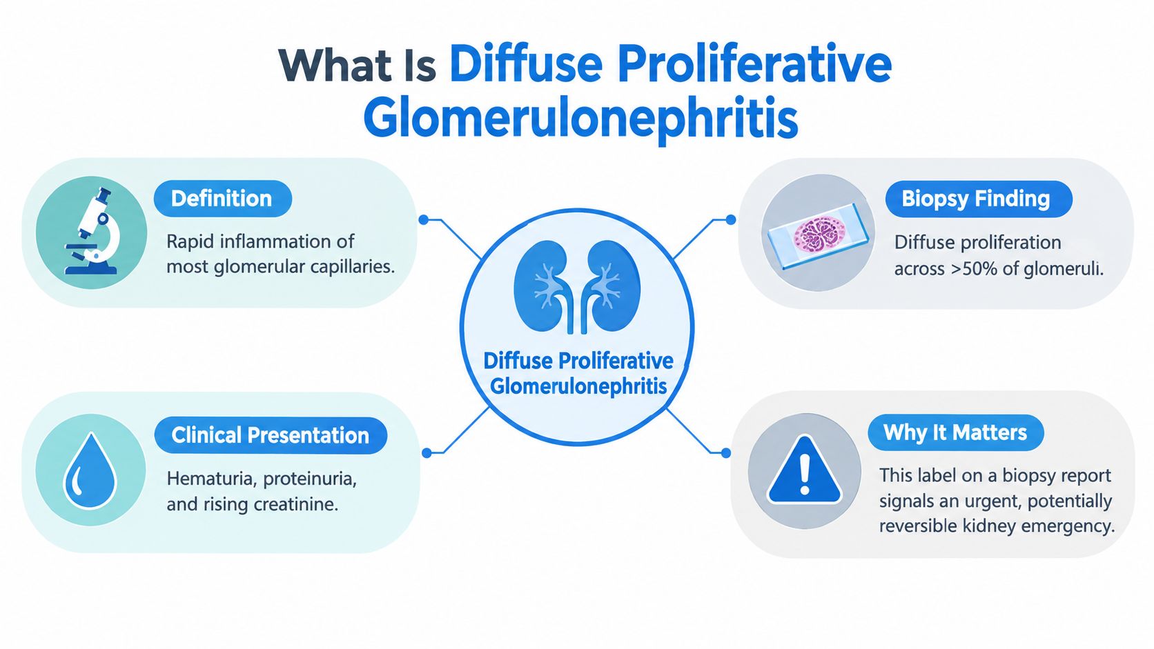
A patient may therefore develop a nephritic presentation, including:
- Hematuria: Red blood cells cross the injured filtration barrier, turning urine pink, red, brown, or tea-colored.
- Proteinuria: The damaged barrier permits more protein to enter the urine.
- Edema: Salt and water retention, combined with urinary protein loss, can produce swelling.
- Hypertension: Reduced filtration and fluid retention increase blood pressure.
- Reduced kidney function: Falling filtration may cause rising creatinine, fatigue, nausea, or reduced urine output.
Practical rule: Treat “diffuse proliferative” as a warning about the intensity and distribution of injury, not as a treatment order.
The pattern matters because it can be severe and potentially reversible, but the window for identifying the cause may be narrow. Residents preparing for nephrology exams may find structured review useful, particularly resources such as Ace Med Boards study strategies, which can help organize pathology, serology, and clinical reasoning around glomerular disease.
Understanding the Pathophysiology and Histology
The glomerular capillary wall normally balances filtration with protection. In DPGN, immune activity disrupts that balance. Immune complexes can deposit in the mesangium or along other parts of the glomerular filtration barrier, activating complement and recruiting inflammatory cells.
Endocapillary hypercellularity is a central microscopic feature. Endothelial cells swell or proliferate, and leukocytes enter the capillary loops. The resulting crowding narrows the lumina through which plasma should flow.
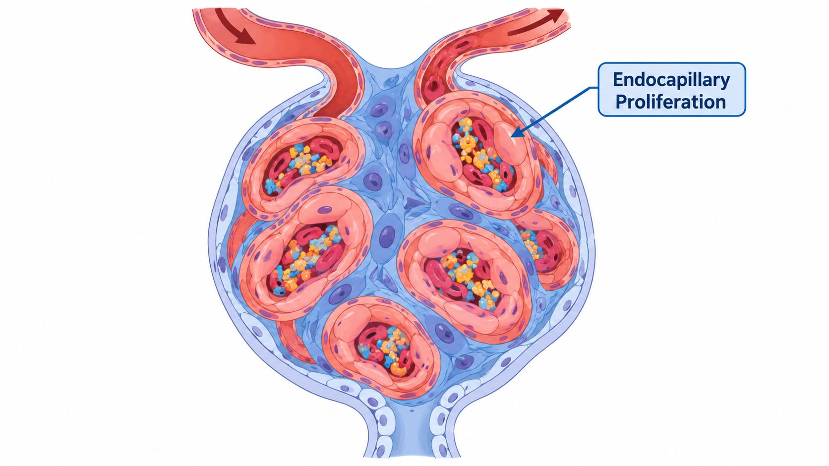
That narrowing explains why kidney function can deteriorate. Less blood reaches the filtering surface, while inflammatory injury makes the barrier more permeable to red blood cells and protein. The patient's urine findings aren't separate problems. They are direct consequences of the altered filter.
Reading the biopsy language
Light microscopy may show:
- Diffuse endocapillary hypercellularity across most glomeruli.
- Leukocyte infiltration, often with neutrophils and mononuclear cells in infection-related disease.
- Narrowed or occluded capillary lumina.
- Crescents or necrosis in more aggressive cases.
- Mesangial and endothelial expansion related to immune activity.
Immunofluorescence adds the immune signature. Infection-related disease commonly produces coarse granular IgG and C3 along capillary walls. Lupus nephritis can show deposits in mesangial, subendothelial, and subepithelial compartments, reflecting a broader immune-complex process.
Electron microscopy provides another layer of confirmation. In postinfectious disease, subepithelial hump-shaped deposits support the diagnosis. The location and character of deposits help distinguish related patterns that may require very different management.
For clinicians building a regular learning routine, ProMed+ Physician CME provides on-demand access to a dynamic clinical curriculum library with over 200 AMA PRA Category 1 Credits™ and content intended to fulfill 100% of state licensure mandates.
A visual review can reinforce the relationship between capillary obstruction, immune deposits, and filtration failure:
Primary Causes and Epidemiological Context
A patient with new hematuria, edema, and rising creatinine may have the same diffuse proliferative pattern on biopsy in two very different diseases. That distinction drives treatment. One pathway is infection-associated disease, where the immediate job is to identify and treat the infection and avoid exposing the patient to harmful immunosuppression. The other is immune-complex autoimmune disease, where delayed immunosuppression can cost kidney function.
Infection-associated disease
Acute post-streptococcal glomerulonephritis follows infection with nephritogenic strains of Streptococcus pyogenes. It remains a classic cause in children, and it still appears where throat or skin infections are not recognized or treated early. The practical clue is timing. A patient improves from the infection, then returns with nephritic features such as hematuria, edema, hypertension, and reduced filtration.
Biopsy does not identify the organism, but it does show the pattern this process tends to produce. In one adult biopsy series, diffuse proliferative and exudative changes appeared in 72% of cases (NCBI Bookshelf). That matters because the tissue can look aggressively inflamed while the management logic remains different from lupus. The resident-level mistake is to react to the word “proliferative” alone. In infection-associated disease, the safer first question is whether an ongoing or recent infection is the driver.
Autoimmune disease
Lupus nephritis creates a different clinical problem. Here, diffuse proliferative glomerulonephritis usually maps to ISN/RPS class IV, defined by involvement of at least 50% of glomeruli. It is then divided into diffuse segmental and diffuse global forms based on how much of each involved glomerular tuft is affected.
That classification is not just descriptive. It helps frame risk, the likely tempo of injury, and the urgency of specialist-directed therapy. In one comparative study, 10-year renal survival was 75% among 35 patients with diffuse proliferative lesions, compared with 52% among 24 patients in the study's diffuse segmental category. The reported relative risk of renal death for diffuse global class IV disease was 1.08, with a 95% confidence interval of 0.68 to 1.70 (comparative lupus nephritis study).

The key clinical pivot is simple. The biopsy pattern shows active glomerular injury, but the cause determines whether you suppress the immune system or step back from that impulse.
Diagnostic Workup and Biopsy Interpretation
Diagnosis works best as a sequence rather than a single test. Start with the clinical story, then use urine studies, kidney function, complement, serology, and biopsy to identify the mechanism.
Start with the syndrome
Urinalysis should look for dysmorphic red cells, red-cell casts, proteinuria, and other sediment abnormalities. Measure serum creatinine and estimate filtration, while checking potassium, bicarbonate, albumin, and blood pressure. Sudden oliguria, pulmonary edema, severe hypertension, or rapidly rising creatinine requires urgent evaluation.
The history should search for recent infection, fever, skin lesions, sore throat, systemic lupus symptoms, medication exposure, endocarditis risk, and features of vasculitis. A normal-looking skin examination doesn't exclude infection-associated glomerulonephritis.
Use serology to narrow the cause
The initial laboratory panel should be directed toward the main competing mechanisms:
- Infection evaluation: Obtain cultures when clinically indicated, review recent antibiotic exposure, and assess streptococcal antibody evidence when post-streptococcal disease is plausible.
- Complement testing: Low complement can support infection-related or immune-complex disease, but it isn't specific enough to establish the cause alone.
- Lupus testing: ANA, anti-dsDNA, and other autoimmune markers should be interpreted alongside systemic findings and complement levels.
- Vasculitis and anti-GBM testing: ANCA and anti-GBM antibodies become important when pulmonary symptoms, rapidly progressive kidney injury, or a crescentic pattern raises concern for those diseases.
- Endocarditis assessment: Persistent fever, murmur, embolic findings, or bacteremia should prompt an infection-focused evaluation before immunosuppression.
Interpret the tissue in three dimensions
Light microscopy identifies the distribution and activity of injury. Look for diffuse endocapillary proliferation, leukocytes, necrosis, crescents, and chronic scarring.
Immunofluorescence determines whether deposits are granular, linear, or sparse. Coarse granular IgG and C3 along capillary walls support postinfectious disease. Lupus nephritis usually shows a more extensive immune-complex pattern.
Electron microscopy can reveal subepithelial hump-shaped deposits in postinfectious glomerulonephritis. The combination of clinical history, complement results, serology, and deposit location is more reliable than any isolated biopsy phrase.
Electrolyte complications can become part of the emergency picture, so clinicians should also review practical resources such as hyperkalemia and potassium management when reduced filtration affects potassium handling.
Treatment Strategies and Clinical Decision Making
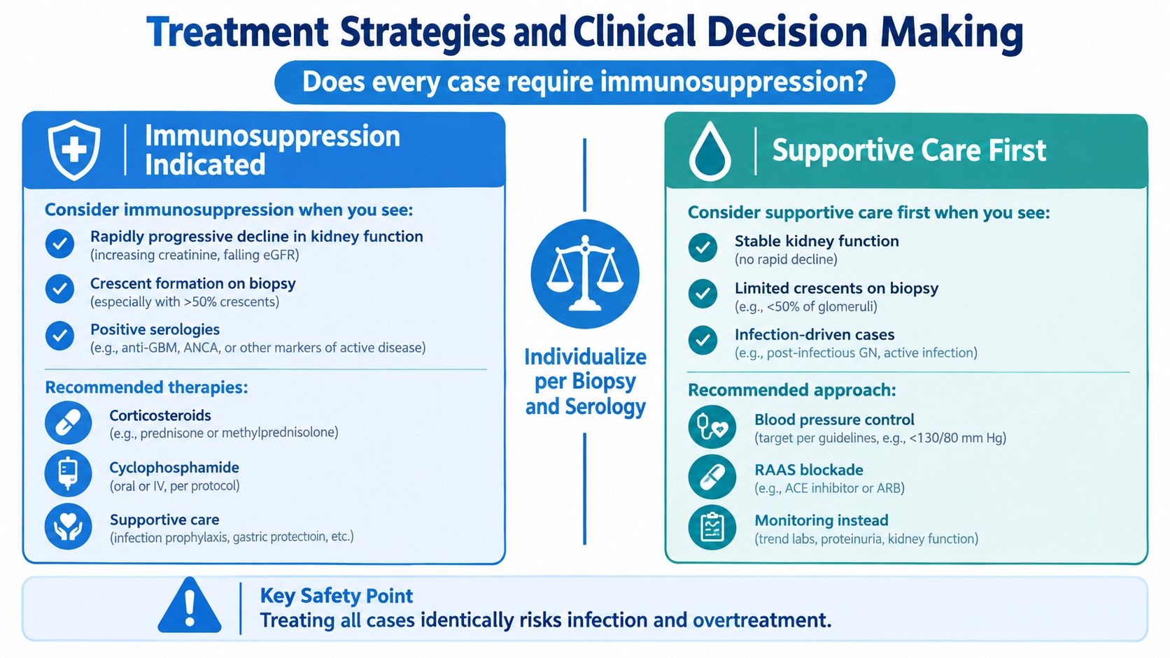
No, every case of diffuse proliferative glomerulonephritis doesn't require immunosuppression. The biopsy pattern describes injury, but infection-associated and autoimmune disease sit on opposite sides of a major safety decision.
For infection-related disease, treatment begins with identifying and eradicating the infection. Supportive care addresses edema, blood pressure, electrolyte abnormalities, and kidney dysfunction. Depending on the clinical situation, this may include fluid and sodium management, diuretic therapy, renal replacement support, and careful medication review.
Immunosuppression can worsen an active infection. This risk is especially important when tuberculosis, hepatitis, parasitic disease, bacterial infection, or endocarditis remains possible.
When immune-directed treatment fits
Active lupus nephritis class III or IV generally requires prompt specialist management. The 2024 KDIGO lupus nephritis guideline recommends glucocorticoids combined with one additional immunosuppressive strategy for active class III or IV disease (KDIGO 2024 guideline).
The specific regimen depends on disease activity, kidney function, comorbidities, pregnancy considerations, infection risk, and biopsy findings. Current approaches may include glucocorticoids with mycophenolate-based treatment, cyclophosphamide, belimumab, or a calcineurin-inhibitor strategy in appropriately selected patients. The decision belongs with nephrology and, when relevant, rheumatology.
When supportive care comes first
A patient with a recent infection, positive cultures, compatible complement findings, and postinfectious biopsy features shouldn't automatically receive steroids because the biopsy says “proliferative.” First control the cause and assess whether kidney function improves.
The difficult exception involves severe rapidly progressive renal failure or persistent inflammatory injury despite infection treatment. A 2025 systematic review of infective-endocarditis-associated glomerulonephritis described improvement after immunosuppressive therapy in 54 cases, worsening in 9, and no change in 3, with substantial mortality (NCBI Bookshelf). These observational findings don't establish a universal protocol. They show why patient selection, infection control, and specialist oversight matter.
Safety principle: Before prescribing immunosuppression, confirm that infection has been reasonably excluded or controlled, and determine whether the biopsy shows active injury, chronic scarring, or both.
Prognosis and Monitoring Response to Therapy
Treatment response should be measured, not guessed from how the patient feels. Edema may improve before the biopsy-level inflammation resolves, while microscopic hematuria can persist even when proteinuria and filtration are moving in the right direction.
For proliferative lupus nephritis, guideline-oriented monitoring uses practical proteinuria milestones. A useful target is approximately 25% proteinuria reduction by 3 months, 50% reduction by 6 months, and a urine protein-to-creatinine ratio below roughly 500 to 700 mg/g by 12 months as a complete-response threshold (guideline-oriented lupus nephritis review).
Build the follow-up around trends
At each review, compare:
- Proteinuria: Is the trajectory improving toward the expected milestones?
- Serum creatinine and eGFR: Is filtration stable, recovering, or declining?
- Urinary sediment: Are active casts and hematuria resolving?
- Complement and serology: Do disease activity markers support response?
- Medication exposure: Is the patient taking therapy consistently and tolerating it?
- Infection status: Could infection explain persistent inflammation or deterioration?
A missed milestone doesn't automatically mean treatment failure. Reassess adherence, drug exposure, the original diagnosis, intercurrent infection, and the balance between active inflammation and irreversible scarring. Persistent hematuria alone also shouldn't trigger automatic escalation when proteinuria, filtration, and serologic activity are improving.
Sudden oliguria, pulmonary edema, severe hypertension, or rapidly rising creatinine is different. Those findings require urgent assessment rather than routine outpatient observation. Acid-base complications may also need focused review, including hydrogen ions and acidosis in kidney disease.
Long-term prognosis depends on the cause, the amount of active versus chronic injury, the response to therapy, and the speed of specialist intervention. In lupus nephritis, the historical survival differences associated with diffuse proliferative categories reinforce the importance of classification and longitudinal monitoring rather than relying on symptoms alone.
Advancing Your Clinical Knowledge Online
A complex diagnosis like DPGN demands continuing education that fits clinical work. Online learning is just as effective as in-person training when the content, assessment, and instructional design are appropriate.
A systematic review of 16 randomized studies involving 5,679 healthcare professionals found little or no difference between e-learning and face-to-face instruction for patient outcomes, professional behaviors, and several skills measures (systematic review of e-learning). Another review identified 11 online CME studies, with 8 of 11, or 72.7%, reporting a significant improvement in at least one outcome, including satisfaction, knowledge, or clinical practice change (online CME review).

Online CE removes travel and fixed classroom schedules while preserving access to structured learning, case review, and assessments. A systematic review of communication-skills training reported that module completion was six times higher among clinicians assigned to online education than among those assigned to face-to-face education.
The industry is shifting toward broader acceptance. Hospitals, clinics, employers, and professional organizations increasingly recognize accredited online CE and certification pathways. The old belief that only AHA or American Red Cross in-person classes are valid isn't true. Validity depends on the relevant employer, licensing board, accreditor, and course requirements, not on physical attendance alone.
For clinicians who want a flexible route to ongoing education, online continuing medical education can support regular study without compromising patient-care responsibilities. ProMed Certifications is one option offering online medical certification courses and continuing education, with self-paced access and certification pathways that include ACLS, BLS, PALS, CPR, and Neonatal Resuscitation.
ProMed Certifications offers online CE and healthcare certification courses that let you study at your own pace while building practical knowledge for clinical decision-making. Visit ProMed Certifications to explore courses that can help you stay current with nephrology-related learning, licensure requirements, and essential certifications.
.avif)
Stay compliant with ProMed+
Certifications included: ACLS, BLS, PALS, CPR & Neonatal Resuscitation
Unlimited continuing education: over 200 hours of accredited CME
All-inclusive: One price. No surprises.
Get certified today

The Advanced Cardiac Life Support (ACLS) is an accredited online medical certification course that teaches medical professionals to respond to nearly all cardiopulmonary emergencies.

The Pediatric Advanced Life Support (PALS) Recertification instructs medical professionals on performing pediatric cardiopulmonary resuscitation in emergencies.

The Basic Life Support (BLS) Recertification is intended to teach healthcare professionals the basic steps of CPR and rescue breathing for adults, children, and infants.

Our online medical certification course for CPR, Automated External Defibrillator (AED), and First Aid is designed to teach adult, child, and infant CPR and AED use.

The Neonatal Resuscitation Certification trains medical professionals in performing resuscitation procedures and life-saving techniques for newborns at the time of delivery.

.png)
.png)
.png)