
Respiratory Distress vs Respiratory Failure: Key Differences
.png)
You are halfway through a busy shift. A patient who looked stable an hour ago is now sitting upright, shoulders tense, breathing faster than before. They can still answer you, but only in short phrases. Their face says effort. Their chest says work. The question is immediate.
Is this respiratory distress, or has the patient crossed into respiratory failure?
New clinicians often get tripped up here because the terms sound similar and can happen in the same patient. At the bedside, though, they are not interchangeable. One describes a body still fighting to keep up. The other means the body is no longer maintaining gas exchange well enough.
That distinction drives everything that follows. It shapes how urgently you escalate oxygen, whether you start noninvasive support, when you call for advanced airway help, and how hard you should push to look beyond a reassuring monitor value. It also shows up constantly in BLS, ACLS, and PALS education because it is one of the most important turning points in emergency care.
The Critical Moment Recognizing Respiratory Compromise
A common bedside mistake is waiting for dramatic collapse before labeling a breathing problem as dangerous.
You see this with the patient who looks “a little short of breath” but is subtly changing position to breathe easier. They lean forward. They pause between words. Their respiratory rate creeps up. In a child, you may see nostril flaring or retractions before the monitor changes much at all.
Those are not small details. They are often the first clues that the patient is spending more and more effort just to move air.
The practical challenge is that respiratory decline is not always loud at first. Early respiratory compromise can look like anxiety, pain, fever, or simple fatigue. That is why experienced clinicians do not ask only, “What is the oxygen saturation?” They ask a broader set of questions.
What the bedside is telling you
Start with what you can see before you touch the patient.
- Body position matters: Patients in trouble often self-position to improve breathing. Tripoding is not random.
- Speech changes matter: Full sentences suggest reserve. Short phrases suggest rising work of breathing.
- Effort matters: Neck muscle use, intercostal retractions, and abdominal breathing are clinical information, not background noise.
A patient can look uncomfortable and still be compensating. That is distress. A patient who is tiring, fading mentally, or failing to oxygenate or ventilate despite support has moved into failure.
Key takeaway: The earlier you recognize increased work of breathing, the more options you have to prevent a crisis.
This is one reason modern certification training matters so much. High-quality online courses can train pattern recognition well when they use realistic scenarios, decision points, and guideline-based case work. For busy clinicians, that format is not a compromise. It is often the most practical way to rehearse the exact bedside judgments that determine whether a patient stabilizes or spirals.
Defining the Spectrum of Respiratory Compromise
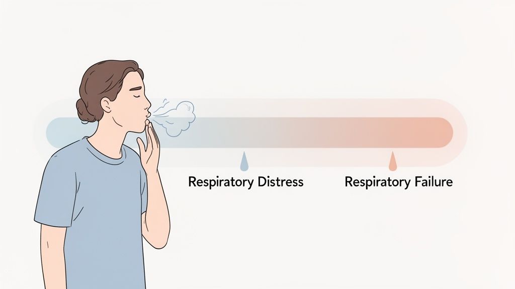
Respiratory distress and respiratory failure sit on the same spectrum. They are not separate islands.
According to a clinical review of respiratory failure vs distress, respiratory distress is the initial clinical presentation of abnormal breathing and can progress to respiratory failure if untreated. That same source makes the key distinction clearly. Respiratory distress describes abnormal breathing patterns, while respiratory failure describes the physiological effects of that abnormal breathing on the body.
What respiratory distress means
Think of respiratory distress as the body in compensation mode.
The patient is working harder to breathe, but they are still managing gas exchange well enough to hold the line. Chemoreceptors sense trouble, neural drive increases, and the respiratory muscles respond with more effort. Clinically, you see tachypnea, retractions, and accessory muscle use.
This is the phase where early action can change the trajectory.
Examples include the patient with early pneumonia who is breathing quickly but is still alert, or the child with bronchiolitis who has visible effort yet remains interactive. The body is under strain, but not yet exhausted.
What respiratory failure means
Respiratory failure begins when compensation is no longer enough.
Now the lungs and respiratory system are not adequately oxygenating, ventilating, or both. The patient is not just working hard. They are losing the physiologic battle.
At this stage, you are no longer focused only on visible effort. You are focused on inadequate gas exchange, rising fatigue, deteriorating mentation, and the need to escalate support quickly.
If you want a deeper review of low-oxygen states that often sit inside this discussion, this primer on hypoxia is useful background.
Why learners get confused
The confusion usually comes from one truth. A patient can be in both.
Someone in respiratory failure may still look distressed. Another patient in respiratory distress may progress rapidly into failure if you miss the warning signs or if the underlying cause worsens. That overlap is normal. It does not make the distinction less useful. It makes the distinction more important.
A way to remember it:
- Distress is the work
- Failure is the consequence
Clinical shortcut: If the body is still compensating, think distress. If oxygenation or ventilation is objectively failing, think failure.
That is why strong training does not stop at memorizing definitions. It teaches you to track the arc of deterioration. Online certification programs do this well when they present cases in sequence, because that mirrors real practice. You rarely meet patients as neat textbook snapshots. You meet them mid-course.
A Side-by-Side Clinical Comparison
At the bedside, you need a clean framework. The easiest way to separate respiratory distress vs respiratory failure is to compare what you see with what the patient’s gas exchange is doing.

Quick comparison table

The objective thresholds that matter
The bedside exam points you in the right direction. The blood gas confirms the physiology.
A 2022 review in Frontiers in Medicine defines Type I respiratory failure as hypoxemic failure with PaO2 below 60 mmHg on FiO2 at or above 0.5, and Type II respiratory failure as hypercapnic failure with PaCO2 above 50 mmHg and pH below 7.35. The same review notes that respiratory distress is a compensatory state where gas exchange is largely preserved, and that the PaO2/FiO2 ratio helps differentiate them. A ratio of 200 to 300 can indicate mild ARDS in failure, while distress typically maintains a ratio above 400 in the absence of mechanical dysfunction, as described in this Frontiers review on respiratory distress and failure.
That gives you a practical ladder:
- Preserved gas exchange with visible effort: distress
- Low PaO2 despite oxygen support: hypoxemic failure
- High PaCO2 with acidemia: hypercapnic failure
- Falling PaO2/FiO2 ratio: worsening oxygenation failure
What this looks like in real patients
A patient with early asthma exacerbation may be breathing fast, using accessory muscles, and still moving enough air to maintain gas exchange. That is distress.
A patient with severe pneumonia who remains hypoxemic despite oxygen, becomes confused, and shows worsening fatigue has moved into failure.
A patient can also transition the other way visually. Early on, they look dramatic because they are fighting hard. Later, as muscles tire, the chest may move less. New clinicians sometimes misread that as improvement. It is often the opposite.
Distress is loud. Failure can get quiet
This is one of the most dangerous misunderstandings in clinical care.
Patients in distress often look busy. Their breathing is fast and forceful. Patients in failure may still look dramatic, but some begin to lose effort as fatigue sets in. Reduced movement in a previously struggling patient is a red flag.
Watch for these turning points:
- Speech drops off: Words become single-word answers or stop altogether.
- Attention drifts: The patient is less engaged, less responsive, or oddly calm.
- Chest movement changes: Less movement after prolonged effort may mean fatigue, not recovery.
- Color worsens: Cyanosis or ashen appearance raises the stakes immediately.
Bedside rule: Never let a single “normal-looking” monitor value override a bad clinical picture.
Why this comparison matters in training
Certification exams test these distinctions because practice depends on them.
A strong online ACLS, PALS, or BLS course can be just as effective as an in-person class for this kind of learning when it uses scenario-based questions and waveform interpretation. The advantage is repetition. You can pause, review, and revisit the exact decision points that separate a patient who needs oxygen and monitoring from one who needs immediate escalation.
Stepwise Assessment and Key Decision Points
When breathing is the problem, a structured exam keeps you from missing the transition from compensation to decompensation.
Use a simple look, listen, and feel sequence. It works fast, and it keeps your attention on the patient instead of only on the monitor.
.png)
Look first
Before you place a stethoscope, inspect the whole patient.
Look for:
- Positioning: Tripoding, neck extension, or refusal to lie flat
- Chest mechanics: Retractions, abdominal breathing, asymmetry
- Facial clues: Nasal flaring in infants, pursed lips in obstructive disease
- General appearance: Anxiety, fatigue, diaphoresis, or fading responsiveness
These findings tell you how hard the patient is working and whether they still have reserve.
Listen with purpose
Now narrow in on the sounds.
Breath sounds can help identify the problem, but they also help you judge severity.
- Wheezing can point toward obstruction.
- Stridor raises concern for upper airway compromise.
- Grunting in infants often signals significant respiratory effort.
- Diminished or absent sounds are especially concerning when the patient also looks tired or confused.
One trap is chasing the sound and missing the patient. Loud wheezing can be serious, but a suddenly quiet chest in a struggling patient can be worse.
Feel for movement and airflow
This step gets overlooked, especially when the room feels rushed.
Assess chest rise, air movement, skin temperature, and whether the effort feels coordinated or weak. If the patient’s effort looks forceful but airflow feels poor, the situation may be deteriorating faster than the pulse oximeter suggests.
Do not overtrust pulse oximetry
One of the most important patient safety issues in this discussion is pulse oximetry bias.
Pulse oximeters can read falsely high oxygen saturation in patients with darker skin tones, which can mask hypoxemia and delay intervention. That means a patient can be moving from respiratory distress into respiratory failure while the SpO2 appears more reassuring than it should. This is why clinicians should combine pulse oximetry with other findings, including mental status and capnography, rather than relying on the monitor alone, as noted in this discussion of pulse oximetry bias and respiratory decline.
Safety point: If the patient looks worse than the pulse oximeter suggests, trust the full clinical picture and reassess aggressively.
That matters even more in patients with rising fatigue, altered behavior, or unexpected drowsiness.
Add capnography when available
Capnography helps when oxygenation data is incomplete or misleading.
If you want a practical refresher, this guide to quantitative waveform capnography explains why it belongs in respiratory assessment, especially when ventilation is the concern.
A good respiratory assessment also benefits from visual review. This video reinforces what worsening respiratory effort can look like at the bedside.
The decision points that change management
Ask these questions in order:
- Is the patient compensating or tiring?
- Is mentation normal, anxious, confused, or fading?
- Is oxygenation preserved, or is hypoxemia breaking through support?
- Is ventilation adequate, or is CO2 retention becoming likely?
- Do the monitor values match the bedside exam?
When those answers stop lining up in a reassuring way, assume the patient is moving closer to failure and escalate.
This is also where online training shines. You can repeat these assessment sequences until they become automatic. For clinicians balancing shifts and recertification deadlines, that flexibility matters. It does not dilute the learning. It supports it.
Immediate Management Strategies
Once you have identified where the patient sits on the spectrum, treatment becomes more targeted. The management of respiratory distress vs respiratory failure is not the same, even when both patients look short of breath.

Manage distress early
Respiratory distress is your chance to interrupt the slide.
The patient is still compensating, so your goal is to reduce work of breathing, support oxygenation, and treat the cause before fatigue sets in.
Practical moves include:
- Positioning: Sit the patient upright if tolerated.
- Low-level oxygen support: A nasal cannula may be enough for mild distress.
- Targeted therapy: Bronchodilators, suctioning, or other cause-specific treatment depending on the scenario.
- Frequent reassessment: Distress is dynamic. Improvement should be visible, not assumed.
The point is not just to “put on oxygen.” It is to lower the physiologic burden before the patient burns through reserve.
Escalate quickly in failure
When the patient is in respiratory failure, oxygen alone may not solve the problem.
A practical teaching point from discussion of oxygen delivery decisions in distress and failure is that a nasal cannula may suffice for mild distress, while respiratory failure often requires high-flow oxygen with a non-rebreather or positive pressure support with NIV. That same source notes an important escalation threshold. If SpO2 remains below 90% or PaO2 remains below 60 mmHg despite initial oxygen therapy, the patient needs a higher level of support.
That gives you a useful sequence:

When NIV makes sense
Noninvasive ventilation can buy time and improve gas exchange in selected patients.
It is especially useful when the patient is still protecting the airway, can cooperate, and needs help with ventilation or oxygenation without immediate intubation. If the patient is becoming obtunded, vomiting, unable to clear secretions, or rapidly deteriorating, NIV may not be enough.
Do not let a temporary response create false comfort. A patient who improves for a few minutes and then tires again is declaring how fragile they are.
Practical tip: Improvement after an intervention should show up in effort, mentation, and overall appearance, not just on the monitor.
Know when to move toward definitive airway management
Intubation is not the first move for every sick respiratory patient. It is the next move when less invasive support is failing or unsafe.
Think seriously about invasive airway support when you see:
- Inability to protect the airway
- Profound hypoxemia despite oxygen or NIV
- Worsening mental status
- Severe fatigue with declining respiratory effort
- Hemodynamic instability in a deteriorating patient
If you need a refresher on escalation choices, this review of advanced airway options and best practices is a helpful companion.
Match the device to the physiology
That is the core management lesson.
Do not treat every dyspneic patient the same. The patient in distress may improve with positioning, a nasal cannula, and disease-specific therapy. The patient in failure may need a non-rebreather, NIV, or a rapid move toward intubation. The wrong pace of escalation is what gets people into trouble.
This is another area where online certification has real value. Device selection, failure thresholds, and escalation logic are highly teachable in digital case scenarios. A clinician can replay a management sequence, compare options, and practice the decision tree until it becomes second nature.
The High Stakes of Unmanaged Respiratory Failure
When respiratory failure is missed or treated too late, the consequences are severe. The most serious form is acute respiratory distress syndrome, or ARDS.
ARDS is the extreme end of this conversation. It reflects severe respiratory failure with substantial morbidity and mortality, even with modern care.
According to the NCBI Bookshelf review on Acute Respiratory Distress Syndrome, ARDS affects up to 15% of ICU admissions. Mortality remains tied to severity, at 27% for mild ARDS, 32% for moderate ARDS, and 45% for severe ARDS.
Why these numbers matter clinically
Those figures are not just exam facts.
They explain why clinicians are taught to react early to increased work of breathing, dropping oxygenation, and changing mental status. Missing the shift from distress to failure does not only delay treatment. It can move the patient closer to a far more dangerous syndrome.
The cascade is often predictable
The progression usually follows a recognizable path:
- Rising work of breathing
- Increasing fatigue
- Worsening oxygenation or ventilation
- Delayed escalation
- More invasive support and more risk
That is why respiratory assessment gets repeated so often in emergency, ICU, pediatric, and transport settings. The patient’s status can change quickly, and when it does, the window for easy correction narrows.
Clinical reality: It is much easier to prevent respiratory failure than to reverse advanced failure after the patient is exhausted.
This is also why respiratory content carries so much weight in certification education. The algorithms matter, but the judgment behind them matters more. Recognize the pattern early, and you can prevent a far worse outcome.
Advance Your Skills with Modern Certification
Differentiating respiratory distress vs respiratory failure is not a niche skill. It is core clinical judgment for nurses, physicians, EMS professionals, respiratory therapists, and anyone who responds to acute deterioration.
You need three things to do it well:
- Pattern recognition
- Objective interpretation
- Decisive escalation
Those skills are built through repetition, not just one-time exposure. That is one reason online certification has become such a strong format for healthcare professionals. It lets you revisit scenarios, pause on difficult concepts, and learn around a real clinical schedule.
The old assumption that only in-person training from a narrow group of providers is valid no longer reflects how healthcare education works. Accredited online certifications are widely used, increasingly recognized, and well suited to the way clinicians maintain competence. For many learners, online education is not only as effective as in-person training. It is a better fit because it improves access, flexibility, and review.
That matters in courses tied to real emergency decisions. If you can repeatedly practice recognizing accessory muscle use, worsening mentation, oxygen delivery choices, and escalation thresholds, you are building the kind of readiness that carries over to patient care.
Good certification training should feel usable on your next shift. It should help you sort out the anxious tachypneic patient from the exhausted failing one. It should help you avoid overreliance on a single monitor. And it should give you confidence that when a patient starts to slide, you will catch it early.
If you are ready to sharpen those bedside decisions and complete your recertification on your schedule, ProMed Certifications offers online ACLS, BLS, PALS, CPR, neonatal, CE, and CME programs built for modern healthcare professionals. The courses are self-paced, guideline-aligned, and designed to deliver the same practical confidence clinicians need in real respiratory emergencies.
.avif)
Stay compliant with ProMed+
Certifications included: ACLS, BLS, PALS, CPR & Neonatal Resuscitation
Unlimited continuing education: over 200 hours of accredited CME
All-inclusive: One price. No surprises.
Get certified today
The Advanced Cardiac Life Support (ACLS) is an accredited online medical certification course that teaches medical professionals to respond to nearly all cardiopulmonary emergencies.
The Pediatric Advanced Life Support (PALS) Recertification instructs medical professionals on performing pediatric cardiopulmonary resuscitation in emergencies.
The Basic Life Support (BLS) Recertification is intended to teach healthcare professionals the basic steps of CPR and rescue breathing for adults, children, and infants.
Our online medical certification course for CPR, Automated External Defibrillator (AED), and First Aid is designed to teach adult, child, and infant CPR and AED use.
The Neonatal Resuscitation Certification trains medical professionals in performing resuscitation procedures and life-saving techniques for newborns at the time of delivery.

.png)
.png)