


Narrow Complex Tachycardia Management
.png)
A patient is sitting upright on the stretcher, saying the heart is racing. The monitor shows a fast rhythm with narrow QRS complexes, while the room starts moving toward the usual SVT checklist. Before reaching for adenosine, pause. Narrow complex tachycardia management begins with the patient, not the monitor.
The key bedside questions are simple but consequential. Is the patient stable? Is the rhythm regular? Is it AVNRT or AVRT, or are you looking at sinus tachycardia, atrial tachycardia, atrial flutter, or atrial fibrillation? This guide walks through those decisions in the order they matter, with practical actions, doses, rhythm clues, and the pitfalls that can change treatment.
First Sixty Seconds at the Bedside
The first minute is not a race to name the rhythm. It's a focused search for instability, reversible threats, and evidence that the monitor is telling the truth.
Start with a rapid ABCDE assessment:
- Airway and breathing: Confirm the patient can speak, assess work of breathing, and provide oxygen when hypoxemia is present.
- Circulation: Check blood pressure, skin temperature, capillary refill, mental status, and the quality of the peripheral pulse.
- Disability: Look specifically for altered mental status, collapse, or new neurologic findings.
- Exposure and context: Search for bleeding, infection, dehydration, medication effects, pregnancy, stimulant use, and other causes of secondary tachycardia.
Place the patient on continuous ECG and oxygen saturation monitoring. Establish two large-bore IVs when the presentation is concerning, and obtain a 12-lead ECG as soon as possible. The 12-lead helps identify the rhythm and ischemic changes, but it must not delay treatment when the patient is clearly unstable.
Put a finger on the pulse while looking at the monitor. A pulse that matches every displayed QRS supports a perfusing rhythm. A mismatch raises concern for artifact, ectopy, or a monitor rate that isn't the effective ventricular rate.
Find the first branch
The immediate decision is stable versus unstable.
Instability includes:
- Hypotension or signs of shock
- Altered mental status
- Ischemic chest pain
- Acute heart failure or pulmonary edema
For an unstable patient, call for help, prepare for synchronized cardioversion, support oxygenation and ventilation, and place defibrillation pads. The 2015 ACC/AHA/HRS guidance recommends synchronized cardioversion when SVT is hemodynamically unstable and vagal maneuvers or adenosine are ineffective or not feasible, as summarized in this guideline document.
If the patient is stable, you have time to inspect the tracing and attempt vagal stimulation. That doesn't mean you can ignore the cause. Severe anemia, dehydration, pulmonary embolism, sepsis, and thyroid storm can all produce a fast narrow rhythm that may worsen if you suppress the rate.
Practical rule: Treat instability immediately, but treat the reason for the tachycardia whenever the rhythm is a response to another illness.
For clinicians maintaining emergency credentials, ACLS Certification from ProMed Certifications is an online option that lets you take the course on your schedule, complete the exam, and receive a card in as quick as one hour or less, according to the provider's catalog information.
Reading the ECG for the Right Diagnosis
A narrow QRS, generally less than 120 milliseconds, tells you that ventricular activation is using the normal conduction system or another pathway that reaches the ventricles quickly. It doesn't tell you which atrial rhythm is driving the heart.
Start with regularity. A regular narrow rhythm suggests sinus tachycardia, AVNRT, AVRT, atrial tachycardia, or atrial flutter with fixed conduction. An irregular rhythm points toward atrial fibrillation, variable atrial flutter, multifocal atrial tachycardia, or atrial activity with changing conduction.
Look for P waves in leads II, V1, and aVL. Retrograde P waves may sit just after the QRS in AVRT or atypical AVNRT. In typical AVNRT, atrial activity can be buried inside the QRS, creating a regular rhythm with no obvious P waves.
The most frequently missed rhythm is typical atrial flutter with 2:1 conduction. The ventricular rate is often around 150 beats per minute, and flutter waves may be hidden inside the T waves. Adenosine can briefly increase AV block and expose those flutter waves without terminating the atrial rhythm. That response is diagnostically useful, but it isn't a successful conversion.
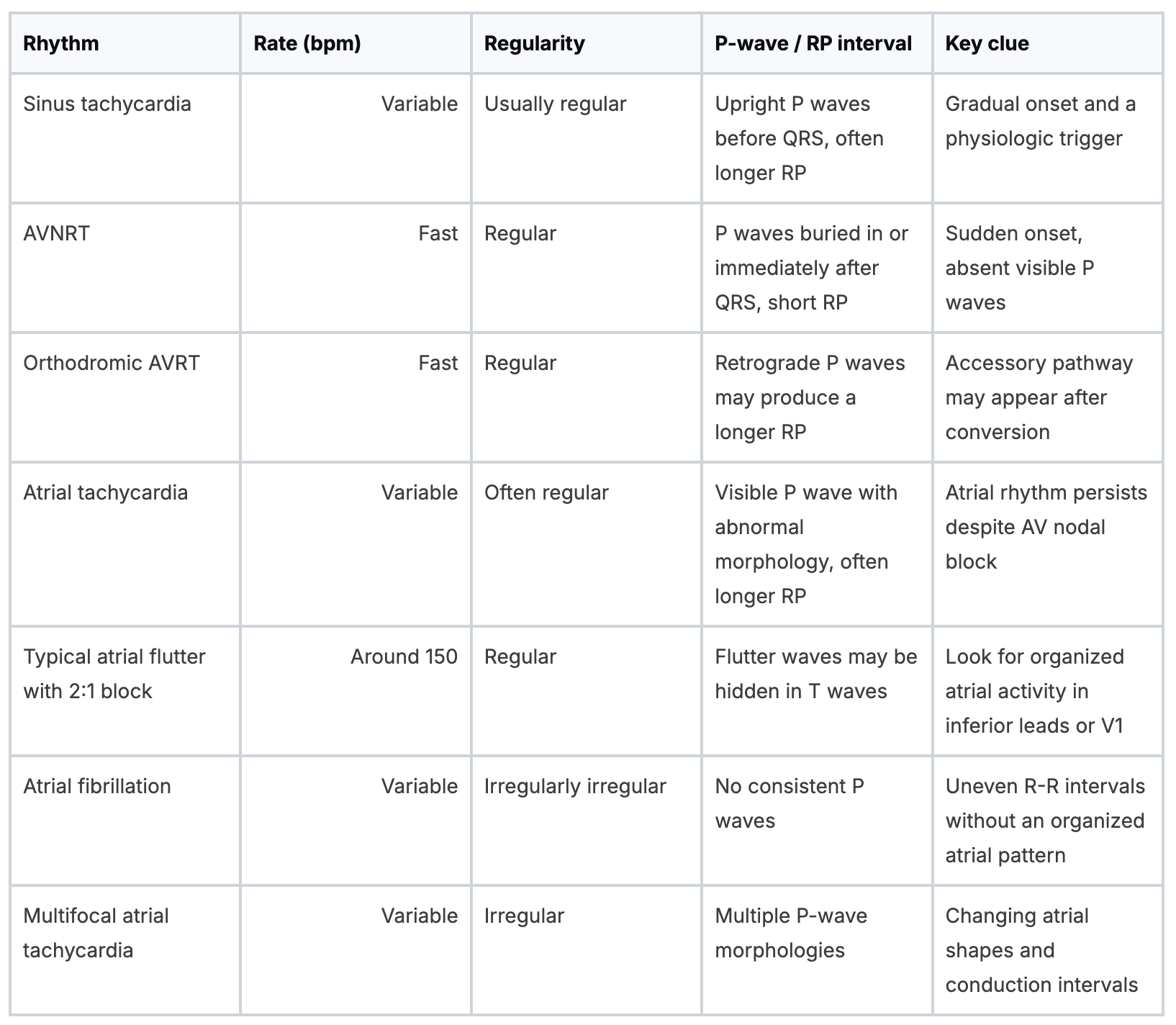
Check lead placement if the atrial activity looks strange. Misplaced precordial or limb leads can create false P-wave patterns, while artifact can mimic atrial fibrillation. A clean rhythm strip may be more useful than a computer label.
For a practical review of waveform recognition and algorithm use, see this basic ACLS ECG interpretation guide.
ECG diagnosis overrides the algorithmic guess. A regular rate around 150 isn't automatically SVT, and an irregular narrow rhythm isn't automatically atrial fibrillation.
Vagal Maneuvers and Adenosine in Practice
For a stable, regular narrow-complex rhythm that appears AV-node dependent, vagal maneuvers come first. They increase parasympathetic influence on the AV node and may interrupt AVNRT or AVRT. They won't reliably terminate atrial flutter or atrial tachycardia, but they can reveal the atrial rhythm.
The modified Valsalva is usually the most practical approach. Position the patient semirecumbently and ask for a forceful strain against a closed glottis for 15 seconds. Immediately lay the patient supine and lift the legs, then reassess the rhythm and pulse. Explain the sequence before starting so the patient doesn't stop during the strain.
Carotid sinus massage is another option in selected adults. Keep the patient supine, use continuous monitoring, and auscultate for a carotid bruit before proceeding. Avoid the maneuver when carotid disease or a bruit makes it unsafe. Don't use eye pressure or face immersion in adults.
.png)
Make adenosine a procedure
If the patient remains stable and the rhythm still looks AV-node dependent, prepare adenosine deliberately. Use a proximal IV site, ideally near the elbow, because the drug's half-life is extremely short. Push it rapidly, then follow immediately with a saline flush.
The common adult sequence is:
- 6 mg rapid IV push, followed by a 20 mL saline flush
- If needed, 12 mg
- If the rhythm persists, a second 12 mg may be given
Warn the patient about flushing, chest discomfort, a sense of impending doom, and a brief pause or asystole. Those sensations are often short-lived, but preparing the patient reduces panic and prevents them from pulling away during administration.
Avoid adenosine in severe asthma, second- or third-degree AV block, or sick sinus syndrome without pacing. Don't use it as the treatment for pre-excited atrial fibrillation in WPW. Continuous rhythm recording matters because adenosine may terminate AVNRT or AVRT, or uncover atrial flutter or atrial tachycardia.
Record a 12-lead during the pause whenever possible. If the rhythm converts and immediately returns, document both events. Recurrent episodes may require a longer-acting agent such as a beta-blocker or calcium channel blocker, provided ventricular function and blood pressure make that choice safe.
This adenosine reference provides additional medication-focused education for clinicians reviewing the drug's role.
Rate Control, Rhythm Control, and Cardioversion
Once the rhythm is identified, decide whether the immediate target is rate, rhythm, or electrical restoration. Rate control can be reasonable for atrial fibrillation or flutter when the patient is stable, while AVNRT and AVRT often call for termination rather than slowing the ventricles.
For rate control, one option is metoprolol 5 mg IV over 2 minutes, repeated every 5 minutes up to 15 mg. Another is diltiazem 0.25 mg/kg IV over 2 minutes. Both can lower blood pressure, and diltiazem can worsen acute heart failure. Reassess after each dose instead of treating the number on the monitor in isolation.
Rhythm-control options require tighter patient selection. Ibutilide 1 mg IV over 10 minutes can be considered in appropriate stable patients, but avoid it when the QT is long or structural heart disease is present. Flecainide may be considered in selected stable patients without coronary disease. These drugs aren't interchangeable, and the rhythm diagnosis must support the choice.
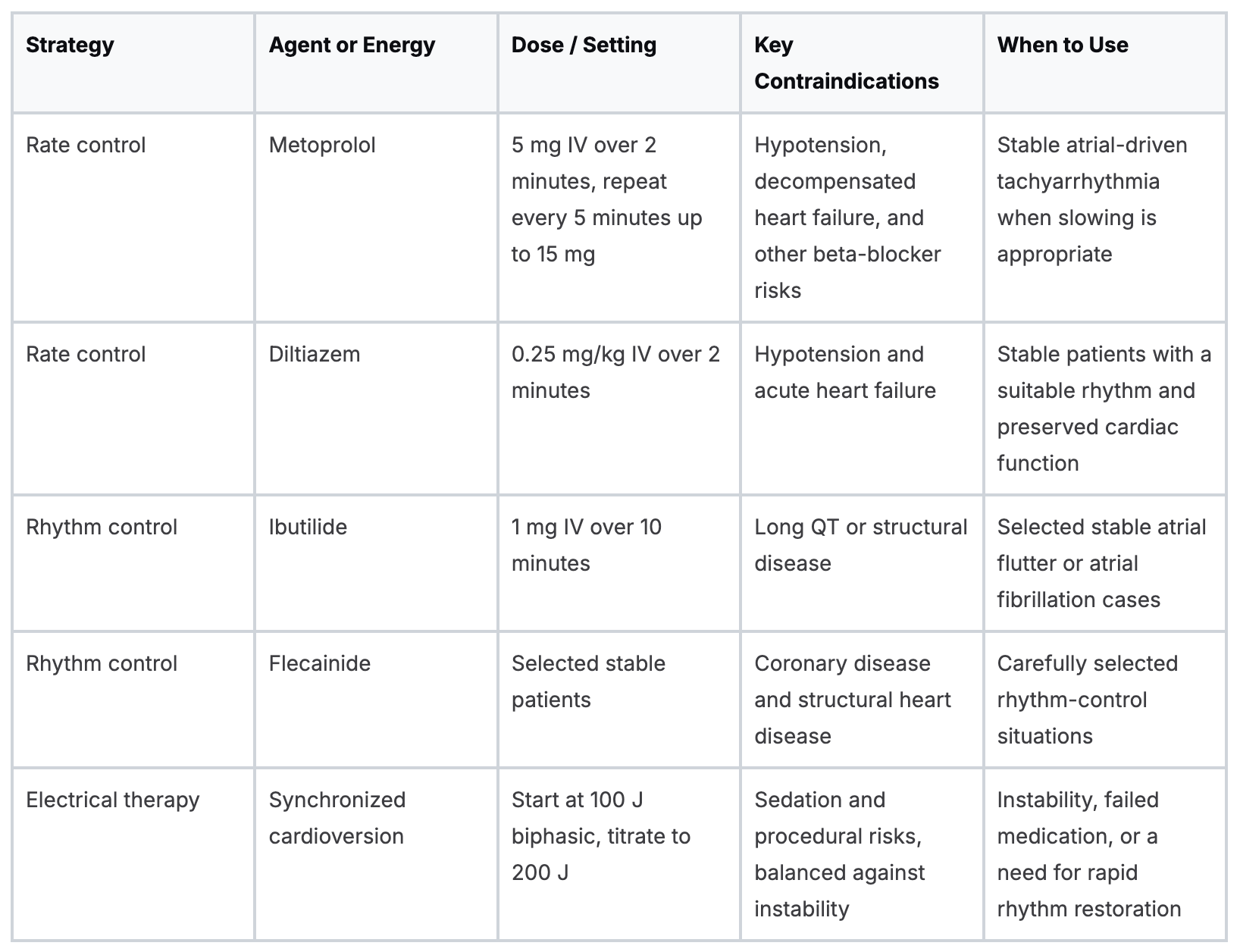
The threshold for electricity is clinical, not emotional. If medications fail, worsen the blood pressure, or delay definitive treatment, synchronized cardioversion becomes the safer choice. The ACLS tachycardia algorithm keeps the central principle clear: unstable patients need synchronized cardioversion rather than a prolonged medication ladder.
Special Situations That Change the Plan
Some presentations look like ordinary SVT until one feature changes the treatment. WPW with atrial fibrillation, pregnancy, pediatric tachycardia, and hemodynamic instability each require a deliberate adjustment.
WPW with atrial fibrillation
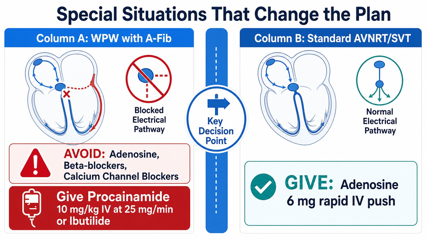
Pre-excited atrial fibrillation can conduct rapidly through an accessory pathway. Avoid AV-nodal blocking drugs, including adenosine, beta-blockers, and calcium channel blockers, because they can favor conduction through the accessory pathway.
For a stable patient, use procainamide 10 mg/kg IV at 25 mg/min or ibutilide, with expert support and continuous monitoring. If instability develops, synchronized cardioversion is the priority. A post-conversion ECG may reveal a short PR interval or delta wave, so don't discard the sinus tracing.
Pregnancy
Begin with vagal maneuvers and use adenosine when the rhythm is appropriate. Adenosine is Category C but has clinical support in pregnancy. If further treatment is needed, metoprolol or diltiazem may be considered with fetal monitoring, while ibutilide should be avoided.
The practical balance is maternal stabilization and fetal safety. Involve obstetric and cardiology teams early when symptoms persist, the diagnosis is uncertain, or electrical therapy becomes necessary.
Pediatrics
Pediatric dosing is weight-based. Give adenosine as a 0.1 mg/kg rapid push, with a maximum of 6 mg for the first dose and 12 mg for the second dose. Diltiazem is generally avoided in children, and synchronized cardioversion starts at 0.5 to 1 J/kg.
Use pediatric pads and equipment, obtain help early, and don't let repeated failed access attempts delay escalation in an unstable child.
Hemodynamic instability
Instability changes the order of operations. Skip the medication ladder, provide airway and circulatory support, sedate when feasible, and deliver synchronized cardioversion at 100 J biphasic, escalating to 200 J.
If synchronization isn't possible and the patient is deteriorating, unsynchronized defibrillation is a last resort. The American Heart Association 2025 ACLS tachycardia algorithm also highlights that energy selection depends on the rhythm subtype, with higher energy emphasized for atrial fibrillation and flutter.
Disposition and Keeping Your ACLS Skills Sharp
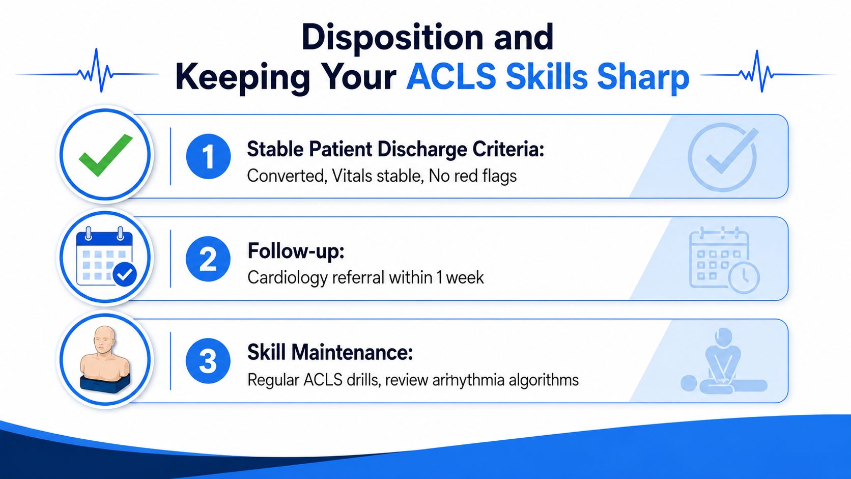
Conversion is not the end of the encounter. Before discharge, confirm that the patient remains stable, symptoms have improved, and no red flags suggest ischemia, heart failure, serious secondary illness, or an unresolved rhythm diagnosis.
A stable patient with converted SVT and no concerning features may be discharged after a brief observation period. Arrange cardiology follow-up, and consider outpatient Holter monitoring or electrophysiology consultation when episodes recur.
Document the event precisely:
- Before conversion: Save the rhythm strip and 12-lead ECG whenever available.
- During treatment: Record the maneuver, exact adenosine dose, IV site, flush, response, and adverse effects.
- After conversion: Save the post-conversion ECG and note whether accessory-pathway features appear.
- After cardioversion: Record the synchronized energy, number of attempts, sedation, complications, and final rhythm.
Disposition also depends on the substrate. Lone AVNRT generally has a benign prognosis, while atrial flutter and atrial tachycardia require evaluation for anticoagulation according to CHA2DS2-VASc and for an underlying cause. Symptomatic WPW warrants electrophysiology referral.
Online learning belongs in skill maintenance
Online CE and certification are now valid, accessible options for healthcare professionals. The idea that only AHA or American Red Cross in-person classes count isn't accurate. Acceptance still depends on employer, credentialing, and regulatory requirements, so verify those details before enrollment, but online learning itself isn't lower quality.
Peer-reviewed evidence supports that position. A systematic review found internet-based e-learning was at least as effective as traditional learning for improving clinician behavior and superior to no instruction, although it found insufficient evidence for patient outcomes because few studies measured them directly. Another review included 59 studies and 6,750 students, finding significantly higher gains online in 12 of 50 knowledge studies and in 6 skills studies, with the authors concluding that online e-learning was equivalent and possibly superior to traditional learning (health professions eLearning review).
A review of evidence-based healthcare education found pure e-learning improved knowledge and skills compared with no learning, while blended learning improved attitudes and behavior compared with face-to-face learning (e-learning review). A 2024 systematic review and meta-analysis also found spaced digital education improved knowledge, skills, confidence, and clinical behavior change in health professionals.
That evidence matches the direction of healthcare education. Hospitals, clinics, and professional organizations are increasingly recognizing accredited online CE and certification pathways. Online formats can reduce travel, protect shift flexibility, and support repeat review without removing the need for serious assessment or employer acceptance checks.
ProMed Certifications offers online ACLS and other healthcare certification courses that let clinicians study around demanding schedules and review rhythm decisions from anywhere. Visit ProMed Certifications to explore an accessible way to keep your ACLS knowledge current and connect narrow complex tachycardia management with practical certification preparation.
.avif)
Stay compliant with ProMed+
Certifications included: ACLS, BLS, PALS, CPR & Neonatal Resuscitation
Unlimited continuing education: over 200 hours of accredited CME
All-inclusive: One price. No surprises.
Get certified today

The Advanced Cardiac Life Support (ACLS) is an accredited online medical certification course that teaches medical professionals to respond to nearly all cardiopulmonary emergencies.

The Pediatric Advanced Life Support (PALS) Recertification instructs medical professionals on performing pediatric cardiopulmonary resuscitation in emergencies.

The Basic Life Support (BLS) Recertification is intended to teach healthcare professionals the basic steps of CPR and rescue breathing for adults, children, and infants.

Our online medical certification course for CPR, Automated External Defibrillator (AED), and First Aid is designed to teach adult, child, and infant CPR and AED use.

The Neonatal Resuscitation Certification trains medical professionals in performing resuscitation procedures and life-saving techniques for newborns at the time of delivery.

.png)
.png)
.png)