

Telemetry Rhythm Interpretation: A Step-by-Step Guide
80%–99% of inpatient telemetry alarms may be false activations, according to evidence discussed alongside the American Heart Association's monitoring practice standards. Most of those alarms do not reflect a rhythm you need to act on, so check the signal and the patient before you name the strip.
A six-step routine gives reference values for each feature. Like a smoke alarm that chirps from a low battery, noise and automated labels can sound urgent while the heart's rhythm is unchanged. Confirm the tracing, then decide whether it changes what you do.
What Telemetry Interpretation Asks You to Do
It's three in the morning. A monitor alarms, but the patient looks comfortable and answers your questions normally. The tracing looks alarming. Do you call an emergency, replace an electrode, or obtain another recording?
Start with the patient, not a rhythm label. Comfort doesn't exclude a significant arrhythmia, but a frightening tracing doesn't establish one either. You need enough clinical and technical information to distinguish those possibilities.
Telemetry continuously monitors the heart's electrical rhythm through a limited set of leads. The tracing appears on a bedside screen, a central monitoring station, or both. Unlike a diagnostic ECG, telemetry emphasizes ongoing surveillance rather than a detailed electrical assessment.
Ask three questions before acting
Is the rhythm real? Decide whether the recording reflects cardiac activity or interference.
What is the rhythm? Describe its rate, timing, atrial activity, and ventricular complexes.
Does it change management now? Connect the finding to symptoms, circulation, and the reason for monitoring.
Think of telemetry as a security camera feed. It helps you notice activity quickly, but you may need a clearer view before deciding what happened. An automated alarm is a prompt to investigate, not a finished diagnosis.
Connect the tracing to the bedside
The bedside relationship matters as much as pattern recognition.
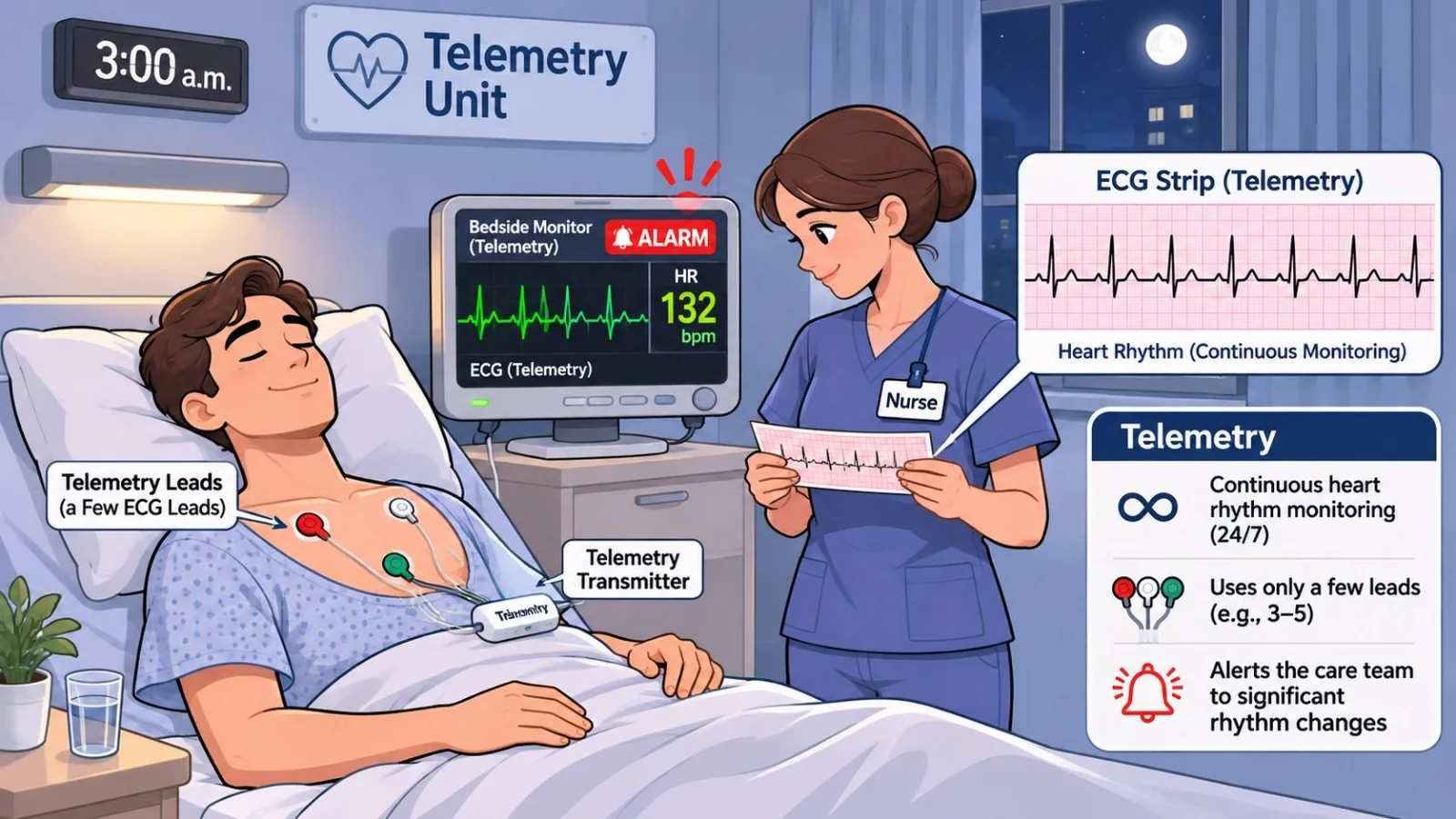
For the comfortable patient, inspect the recording while checking their condition and monitoring connections. For an unresponsive patient without a pulse, activate the emergency response rather than polishing your interpretation.
Practical rule: A rhythm name is useful only when it helps you make the right bedside decision.
Use a consistent reading order, compare measurements with adult reference ranges, and learn the distinguishing clues of common rhythms. Pair those skills with artifact troubleshooting, a clear escalation pathway, and careful review of automated alerts. Each task supports the same outcome: recognizing clinically meaningful changes without treating the monitor instead of the patient.
A Six-Step Routine With Reference Values for Every Strip
A dependable routine reduces the temptation to recognize a familiar shape and stop looking. Use ECG calipers, an electronic measurement tool, or a clean strip and straightedge. Check the recording's time scale before calculating rate or measuring intervals.
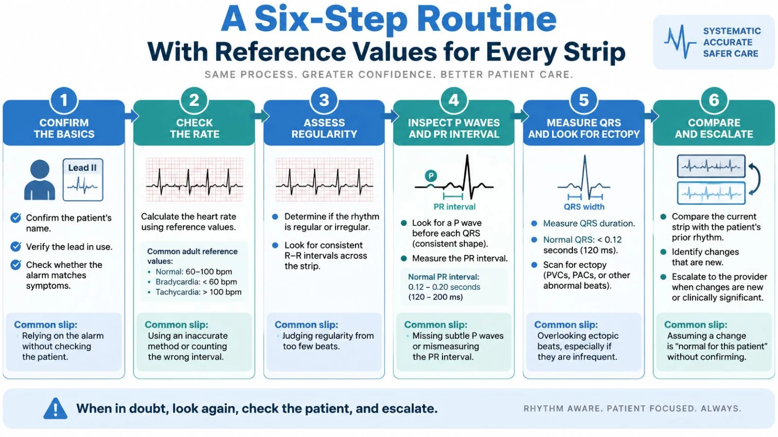
The visual sequence below provides a repeatable order for your review.
Begin with identity, rate, and timing
Confirm the patient and the recording. Match the displayed name with the patient, identify the lead, and check whether the alarm corresponds with symptoms. Inspect several beats rather than a single frozen complex. The common slip is assuming the correct name and a recognizable waveform guarantee a trustworthy recording.
Measure the rate. On a six-second strip, count the R waves and multiply by ten to estimate beats per minute. For a regular rhythm, you can use the large-box method after confirming the recording speed. Don't apply a regular-rhythm shortcut to an irregular tracing, and don't count artifact spikes as ventricular beats.
Assess regularity. Compare consecutive R-R intervals, the spaces between ventricular complexes. Describe the pattern as regular, regularly irregular, or irregularly irregular. A repeating early-beat pattern differs from unpredictably changing intervals, even though both are irregular.
Rate tells you how often the ventricles activate. Regularity tells you how consistently that happens. Neither feature, by itself, establishes the rhythm's origin.
For example, a fast, regular rhythm could be sinus tachycardia or another supraventricular rhythm. You still need to examine atrial activity and ventricular conduction before naming it.
Examine atrial activity and ventricular conduction
Inspect P waves and the PR interval. Look for atrial activity before each QRS, then ask whether every P wave conducts to a ventricular complex. Measure from the beginning of the P wave to the beginning of the QRS. A common mistake is noticing that every QRS has a P wave but overlooking additional P waves that don't conduct.
Also check whether P waves look alike and whether the PR interval stays steady. If you can't see atrial activity clearly, select a more informative lead rather than declaring it absent.
Measure QRS width and inspect unusual beats. Measure the entire ventricular complex, not just its tallest deflection. A narrow QRS generally suggests activation through the usual ventricular conduction system. A wide QRS requires consideration of ventricular origin, aberrant conduction, pacing, or an established conduction abnormality.
Compare an early, differently shaped beat with the surrounding complexes. Don't let an isolated ectopic beat determine your description of the entire underlying rhythm.
Describe, name, and choose the next action. Compare the strip with the patient's prior rhythm. Then decide whether to document the finding, obtain a diagnostic ECG, seek expert review, or escalate immediately. The common slip is treating the monitor's printed label as confirmation.
Adult reference values for rhythm features
Use these adult teaching references alongside your unit's ECG measurement standards. Telemetry is especially useful for rhythm surveillance; clinically important interval abnormalities deserve diagnostic confirmation.
Feature | Normal adult value | What a deviation can suggest |
|---|---|---|
Heart rate | 60-100 beats per minute at rest | Under 60: bradycardia; over 100: tachycardia |
Regularity | R-R intervals steady within a small margin | Irregular pattern: atrial fibrillation or frequent ectopy |
P wave | Upright and present before each QRS in lead II | Absent or abnormal: atrial fibrillation, junctional or ectopic rhythm |
PR interval | Within the adult ECG reference range, consistent from beat to beat | Long: first-degree block; short: pre-excitation or junctional rhythm |
QRS duration | Narrow, within the adult ECG reference range | Wide: ventricular or aberrant origin |
QTc interval | Within the sex-specific reference range for the correction method used | Prolonged: drug effect, electrolyte disturbance or congenital cause |
The QT interval includes ventricular activation and recovery. QTc adjusts that interval for heart rate, so it isn't interchangeable with an uncorrected QT measurement. Check the correction method and the underlying tracing before accepting a displayed QTc.
Finish by describing the features aloud: the rate category, regularity, P-wave relationship, and QRS width. For example: “The rhythm is slow and regular, with a P wave before each narrow QRS.”
That sentence makes your reasoning visible. If you can describe the features but can't confidently name the rhythm, document the objective findings and request review. An accurate description is safer than a confident guess.
Common Telemetry Rhythms and How Urgent They Are
Two strips can both look fast and still call for different responses. Sinus tachycardia often tracks pain, fever, or another stress the body is answering. A sudden regular tachycardia may start from a different site and behave differently. Read the waveform and the patient together before you name the rhythm. Many monitor alarms will not change what you do at the bedside, so confirm the signal and the person in the bed first.
Group rhythms by where activation begins: the sinus node, another supraventricular site, or the ventricles. Conduction blocks form their own group because the problem is transmission between atria and ventricles.
Common telemetry rhythms at a glance
Rhythm | Rate and regularity | Key clue on the strip | Typical urgency |
|---|---|---|---|
Normal sinus rhythm | 60-100 per minute, regular | P before every narrow QRS | Routine |
Sinus bradycardia | Under 60 per minute, regular | P before every narrow QRS, slow | Assess symptoms and pulse |
Sinus tachycardia | Over 100 per minute, regular | P before every narrow QRS, fast | Find the cause |
Atrial fibrillation | Changing rate, irregularly irregular | No consistent P waves, usually narrow QRS | Assess rate and stability |
Supraventricular tachycardia | Fast and usually regular | Narrow QRS, P waves hidden or inverted | Urgent if unstable |
Ventricular tachycardia | Often 100+ per minute, regular or nearly regular | Wide QRS, possible AV dissociation | Emergency if unstable or pulseless |
Ventricular fibrillation | No organized rate | Chaotic baseline with no QRS complexes | Emergency, begin resuscitation |
Asystole | No electrical activity | Flat line confirmed in two leads | Emergency, begin resuscitation |
Second-degree Mobitz II | Atrial rate steady, ventricular rate slowed | Intermittent dropped QRS, fixed PR in conducted beats | Urgent, assess for pacing |
Premature ventricular contractions | Underlying rhythm, irregular only at the early beat | Wide early beat with no preceding conducted P wave | Routine if isolated; review if frequent |
Local protocols, clinical assessment, and current ACLS algorithms set the actual response. Use the urgency column as a teaching map after you have matched the strip to a pulse and a clinical picture.
The chart provides a visual comparison of the patterns.
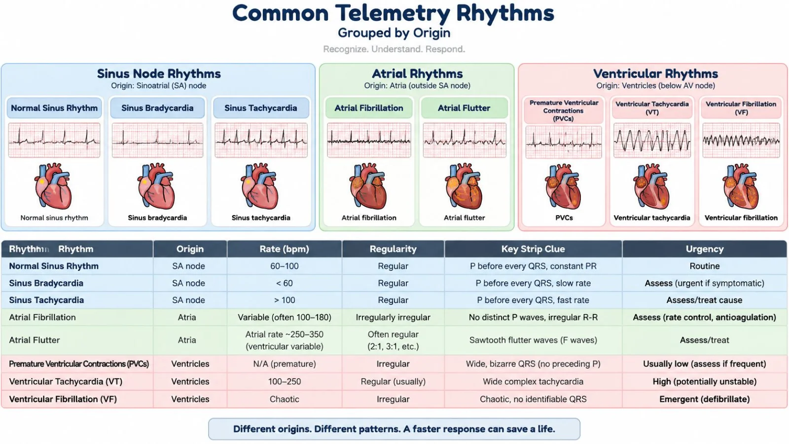
Use clues without turning them into absolutes
Premature ventricular contractions, or PVCs, are a familiar ectopic finding on telemetry. An isolated PVC in an otherwise stable patient often calls for documentation and context. Frequent PVCs, new patterns, or consecutive ventricular beats warrant review. One early wide beat works like a single skipped step on a staircase: note it, then watch whether the next steps stay orderly.
For narrow complexes, start with the usual ventricular pathway, then ask whether P waves lead the complexes. For wide complexes, consider a ventricular origin early. Width is one clue. Pair it with the P-wave relationship and the patient's pulse, because a supraventricular impulse can conduct abnormally and still look broad.
Mobitz II deserves particular attention. A conducted beat can look reassuring and still hide dropped ventricular responses. Our telemetry strip interpretation for nurses resource examines that pattern more closely.
Connecting recognition with response is the aim of ACLS Certification. ProMed's ACLS course is an accredited online medical certification that teaches medical professionals to respond to nearly all cardiopulmonary emergencies.
Finish the feature checks, confirm that the monitor matches the patient, and then attach a name. The rhythm label records those observations so the next clinician can follow the same path.
Separating Real Rhythms From Artifact and False Alarms
The monitor may have lost the signal while the heart's electrical activity continues. A flat trace works like a headphone plug that has slipped halfway out: the recording is still there, and the silence is in the connection. Check the patient and the leads before you call that silence asystole. Dismissing an unfamiliar waveform as artifact can also delay recognition of a true emergency.
An AHRQ patient-safety review described 381,560 audible alarms across 77 ICU beds in 31 days, about 187 alarms per bed per day, and reported that 88.8% of 12,671 analyzed arrhythmia alarms were false, with low-voltage QRS complexes contributing to false asystole alarms when the monitoring lead failed to detect them.
Most of those alarms are not actionable. Confirm the signal, then the patient, and only then name the rhythm.
Recognize the sources of distortion
Loose or dried electrodes, patient movement, muscle tremor, a disconnected lead, and electrical interference all distort the tracing. A wandering baseline drifts when the patient shifts, which makes interval measurements harder and can hide atrial activity the way a wrinkled page hides a line of print.
The checklist below connects a distorted tracing with a cause you can often correct.
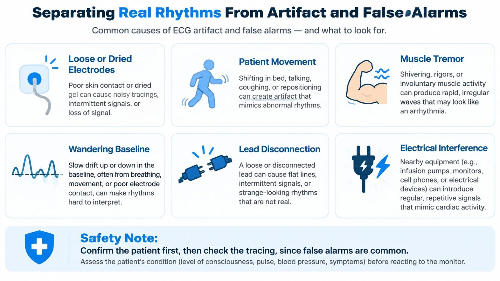
Use a two-stage check. Do not wait to finish inspecting cables before you look at the patient.
Technical validation: Inspect electrode adhesion, anatomical placement, connections, and signal quality. Check whether an apparent pause appears in another lead.
Clinical validation: Assess consciousness, pulse, blood pressure, symptoms, and whether the waveform fits the patient's condition.
Lead selection: When complexes are too small for reliable detection, select a lead with taller QRS complexes and clearer atrial activity. Confirm that the monitor recognizes the beats after the change.
Our guide to avoiding misread rhythms in emergency care provides additional troubleshooting context.
Separate symptom education from rhythm confirmation
A patient may ask whether coffee explains their palpitations. Optimal Native's Spanish-language explicación del café y ritmo cardíaco offers background for that conversation, but it does not replace ECG assessment or establish the cause of an alarm.
Do not silence uncertainty by choosing a rhythm label. Resolve the uncertainty with a better signal and a bedside assessment.
If the patient is well and the recording is noisy, correct the technical problem and reassess. Document the alarm, your assessment, what you changed, and the resulting rhythm. If the patient is deteriorating, seek immediate help while the team verifies the tracing.
When to Escalate and Confirm With a 12-Lead ECG
Telemetry is a screening tool, not a complete diagnostic ECG. A suspicious finding in a stable patient often needs confirmation before rhythm-specific treatment. In an unstable or pulseless patient, confirmation must not delay emergency care.
ACC/AHA electrocardiography competency guidance emphasizes technically adequate acquisition and the risk of incorrect diagnoses and unnecessary treatment when artifact goes unrecognized. The practical lesson is straightforward: obtain a recording you can trust, then connect it to the clinical assessment.
Escalation actions for common telemetry findings
Finding on telemetry | Confirm first | Escalate when |
|---|---|---|
Bradycardia or suspected conduction disease | 12-lead ECG to confirm rate, PR and QRS | Symptoms, hypotension or a high-grade block |
Stable wide-complex tachycardia | 12-lead ECG during the tachycardia | Always assess promptly; follow ACLS if unstable |
Suspected ventricular tachycardia criteria, including AV dissociation or concerning precordial morphology | 12-lead ECG and rhythm review | Manage suspected ventricular tachycardia cautiously pending expert assessment |
Apparent asystole or pause on telemetry | Check connections and signal in a second lead while assessing the patient | Immediately if the patient is unresponsive or pulseless |
Ventricular fibrillation or pulseless rhythm | No confirmation delay | Immediately under ACLS |
Prolonged QTc or suspected electrolyte change | 12-lead ECG and medication review | Notify the provider under local policy; escalate urgently for instability |
For bradycardia or suspected conduction disease, a diagnostic ECG helps establish the relationship between atrial activity and ventricular conduction. It also supports more reliable assessment of PR, QRS, and QTc intervals.
When electrolyte disturbance is suspected, don't interpret the waveform in isolation. Communicate the clinical concern so the responsible clinician can coordinate laboratory evaluation, medication review, and treatment.
Capture the abnormality while it's present
For stable wide-complex tachycardia, obtain a diagnostic ECG during the episode whenever feasible. A recording after termination may not preserve the features needed to identify the mechanism.
Features supporting ventricular tachycardia include atrioventricular dissociation, in which atria and ventricles activate independently, and concerning patterns across the chest leads. These require a diagnostic tracing and experienced review, not just a single telemetry lead.
Preserve the event: Save the onset, representative rhythm, and termination when available.
Give a useful handoff: Report symptoms, pulse, blood pressure, rhythm features, and whether the change is new.
Assign responsibility: Confirm who is obtaining the ECG, contacting the clinician, and continuing bedside observation.
For a narrow-complex differential, the SVT resources from ProMed Certifications can support learning about rhythm assessment and response pathways.
Know your unit's escalation chain before you need it. Identify the bedside responder, responsible clinician, and emergency activation process. During transfers and handoffs, explicitly confirm who owns monitoring and whether the receiving team has a usable signal.
Using AI Rhythm Alerts as a Second Reader
An AI alert can draw attention to an overlooked event. It can also be wrong. The safest role for automated interpretation is a second reader, with clinicians responsible for reconciling its output with the waveform and patient assessment.
In a 2025 Nature Medicine study of 14,606 ambulatory ECG recordings, AI sensitivity for critical arrhythmias was 98.6% versus 80.3% for certified technicians, with false negatives of 3.2 versus 44.3 per 1,000 patients, respectively. Those findings are encouraging, but ambulatory ECG performance doesn't automatically establish performance in every inpatient telemetry setting.
Challenge the label with observable evidence
An automated result should leave you able to inspect the underlying event. A label without an accessible waveform doesn't give you enough information to verify rhythm features or identify interference.
Check the input: Is the recording technically adequate, or is the algorithm classifying noise?
Review the waveform: Can you identify the rate, regularity, atrial activity, and QRS pattern yourself?
Look for disagreement: Does the label conflict with symptoms, circulation, or the visible strip?
Resolve responsibility: Follow local policy for review, escalation, and documentation of disputed alerts.
Limited external validation, difficult-to-explain outputs, workflow barriers, and inconsistent clinician education remain important implementation concerns. A strong result from one system doesn't eliminate the need to evaluate how a particular tool functions in your clinical environment.
Give symptoms priority over reassurance
Say the software identifies no significant abnormality, but the patient reports new dizziness and looks unwell. Don't use the automated result to dismiss the complaint. Assess the patient and obtain appropriate clinical review.
When evidence conflicts: A symptomatic patient takes priority over a reassuring automated label.
The opposite disagreement matters too. An alarming label with a questionable waveform requires review, not automatic treatment. Check every AI alert against the strip and the patient before deciding what it means.
Build the Routine Into Your Next Shift
Poor ECG rhythm identification fell from 46.9% to 5.8% among 76 nurses after an eight-week interactive online program, according to a 2025 International Journal of Nursing Sciences study. The gain came from practice people could repeat, not from one long class.
A weekly strip session works like a code-cart check. You open the same compartments in the same order, so a missing item is obvious before you need it.
Judge the course by evidence you can check
A Cochrane systematic review found little or no difference in several patient outcomes between interactive e-learning and traditional education for licensed health professionals. A separate review of postregistration medical education reported comparable effectiveness and noted limits in the quality of that evidence. Accreditation, course requirements, and employer acceptance are the details worth confirming before you enroll.
A national survey of clinicians documented demand for online courses learners could revisit and fit around work. Self-paced modules let you pause a confusing QRS, replay the strip, and answer again without leaving the unit.
Make practice deliberate
Read five strips weekly: Include clean, noisy, and ambiguous examples, and finish your read before you check the label.
Log each error: Note whether the miss was signal quality, timing, atrial activity, or the patient's symptoms.
Review monthly: Group the repeated misses and run those strips again until the error changes.
On your next shift, take one alarm you would usually silence. Write one line: how the patient looks, whether you can feel a pulse, and whether the waveform is readable enough to name. That line is the drill. It makes you confirm the signal and the person before you accept a rhythm name.
ProMed Certifications offers accredited online courses that connect rhythm recognition with emergency-response decisions. The ACLS course is an accredited online medical certification that teaches medical professionals to respond to nearly all cardiopulmonary emergencies. Its ACLS, BLS, PALS, Neonatal Resuscitation, and CPR courses are based on the latest published cognitive guidelines endorsed by the American Heart Association, and its certification protocols are AHA-aligned. Visit ProMed Certifications for self-paced training you can pause and retake around a shift.