

What Are ECG Artifacts: A Practical Guide to Recognition

ECG interpretation is a critical component of safe emergency and bedside care. Yet even experienced clinicians can hesitate when a monitor suddenly displays a chaotic rhythm, a drifting baseline, or apparent ventricular tachycardia. This guide will help you understand what ECG artifacts are, recognize their patterns, troubleshoot them at the bedside, and protect real clinical decisions from both noisy signals and overconfident digital fixes.
When a Noisy Strip Steals the Story
A telemetry alarm sounds during a busy shift. The monitor shows a fast, wide-complex rhythm that resembles ventricular tachycardia. In the room, however, the patient is awake, speaking clearly, and has a steady pulse. The change began while an arm moved, a blanket was adjusted, or a hand reached for the call button.
The patient may be stable even when the tracing is not.
That mismatch can prompt unnecessary defibrillation, adenosine, or antiarrhythmic medication. The reverse mistake carries its own danger: dismissing a true arrhythmia as artifact, or allowing noise to conceal ischemic changes. Artifact recognition is therefore a frontline ACLS and PALS skill, not a minor technical task.
Practical rule: Correlate before you interpret. Assess the patient, check the pulse and vital signs, then decide whether the monitor's rhythm fits the bedside findings.
Monitoring studies have repeatedly documented artifact-driven misinterpretation, including supraventricular rhythms mistaken for ventricular tachycardia. A noisy strip deserves clinical correlation before treatment, especially when the displayed rhythm conflicts with the patient's appearance and pulse.
Use a simple sequence at the bedside. Define what has changed in the signal, then examine its visual pattern. Correlate that pattern with the patient, inspect the electrodes and cables, correct the likely source, and repeat the tracing. Only after that should you judge whether filtering or software has clarified the record. Denoising can make a questionable signal look convincing, while AI may smooth away clues that would have prompted a fresh assessment. These tools should supplement bedside judgment, not replace it.
What ECG Artifacts Are
ECG artifacts are noncardiac electrical signals or waveform distortions superimposed on the heart's true electrical activity. They can enter the recording through the patient, the electrode and skin interface, cables, acquisition electronics, or electromagnetic activity nearby.
An ECG works like a microphone recording a conversation. The heart provides the intended voice, while movement, muscle tension, loose electrodes, and electrical equipment add room noise. The voice remains present, yet noise can hide words, create false sounds, or make a steady conversation appear chaotic.
Artifact does not by itself indicate heart disease. It indicates that the recorded signal may not accurately represent cardiac activity. Patient movement, skeletal-muscle activity, poor skin preparation, electrical interference, respiration-related baseline movement, and incorrect electrode placement are common sources. A review of electrocardiography found electrode-placement errors in approximately 0.4% to 4% of ECGs, showing that placement problems can distort diagnosis. The electrocardiography artifact review discusses this problem in clinical context.
Start with the tracing and the patient
A genuine cardiac rhythm should fit the patient's physiology. QRS complexes, the palpable pulse, symptoms, blood pressure, and level of consciousness should form a compatible story. Artifact may appear in one lead, affect several leads at once, or produce a pattern that does not follow the expected cardiac cycle.
Use this sequence at the bedside:
- Look at the patient: Is the patient conscious, speaking, perfusing, and showing symptoms that fit the apparent rhythm?
- Inspect multiple leads: A true cardiac event should have a plausible relationship across leads viewing the same electrical activity.
- Check the equipment: Examine electrode contact, cable tension, placement, and signal quality.
- Repeat the recording: If the tracing conflicts with the clinical picture, correct the suspected source and obtain a cleaner strip.
For clinicians reviewing emergency rhythms, ACLS Certification is one example of an online certification pathway offering coursework and an exam around a busy schedule. It does not replace clinical judgment. Structured practice can reinforce the habit of correlating an alarming strip with the patient before acting.
The Four Families of Tracing Noise
Frequency gives you a useful first clue. Ask, “Is this noise slow, fast, regular, or sudden?” The answer often points toward both the source and the correction.
Baseline wander occupies the low-frequency end. It appears as a gradual upward or downward movement of the isoelectric line. Respiration, slow patient movement, electrode drift, and unstable skin contact commonly contribute to it. Because the baseline moves over several beats, it can make ST-segment assessment difficult.
Muscle and motion artifact usually contains higher-frequency or broadband activity. Skeletal-muscle contraction, tremor, shivering, anxiety-related tension, Parkinsonism, and cable movement can create irregular fuzz or rapid spikes. The noise may look disorganized, but the underlying QRS rhythm can remain regular.
Power-line interference is more regular. It typically appears as a 50 or 60 Hz signal, depending on the electrical grid. Nearby equipment, poor grounding, or electromagnetic coupling can produce a fine, repetitive oscillation that looks too uniform to be biological.
Electrode-related artifact crosses the frequency categories. Poor skin preparation, dried gel, loose contact, cable tension, and incorrect placement can create baseline shifts, abrupt deflections, or intermittent signal loss. Electrode problems often become worse when the patient moves.
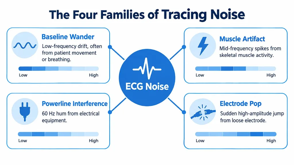
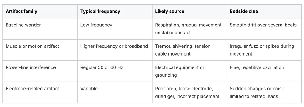
This taxonomy prevents a common mistake: treating every noisy tracing as the same problem. A slow drift needs a different response from a regular electrical hum. A tremor requires a different response from a lifting electrode.
Frequency doesn't diagnose the patient. It helps you locate the problem.
Reading the Pattern on the Strip
A noisy strip becomes easier to interpret when you describe what you see before naming the rhythm. Start with the shape, speed, regularity, and distribution of the abnormal signal.
Baseline wander looks like a slow wave carrying the tracing upward or downward. It may follow breathing or gradual movement and can obscure the ST segment without creating sharp, independent complexes. Muscle artifact appears more like irregular fuzz, often in clusters while the patient shivers, grips, trembles, or shifts position. Tremor can be surprisingly regular and may mimic atrial flutter or ventricular tachycardia.
Power-line interference is different. The oscillation is fine and repetitive, often appearing as a consistent texture over the baseline. Electrode contact problems may create abrupt vertical changes, intermittent loss of signal, or distortion limited to leads connected to the affected electrode.
Three bedside checks
- Compare several leads. Look for the underlying QRS complexes in leads that remain clearer. If the apparent rhythm appears only in one area or doesn't have a convincing multi-lead pattern, question it.
- Check the pulse and symptoms. A patient with a stable pulse and no symptoms deserves reassessment before treatment for a monitor-only ventricular rhythm.
- Look for simultaneous contamination. Noise affecting several leads at the same moment may reflect movement, grounding, or an environmental source rather than a new cardiac event.
The rhythm can also reveal the answer. If the baseline looks chaotic but the visible R-R intervals remain regular, muscle artifact is more plausible than atrial fibrillation. If the monitor suggests a lethal rhythm while the patient remains alert with a corresponding pulse, artifact becomes the leading technical concern.
For additional foundational practice, this guide to reading ECGs can help learners build a systematic approach to rate, rhythm, intervals, and waveform interpretation.
Why Artifact Recognition Protects Real Patients
Artifact can imitate a dangerous rhythm and prompt treatment the patient does not need. It can also obscure a real abnormality, creating false reassurance and delaying care. The same noisy tracing can therefore produce opposite clinical errors.
The ambulatory study included 1,000 recordings and found artifacts more often in telemetry than Holter recordings, with 28 of 500, or 5.6%, telemetry recordings affected compared with 20 of 500, or 4.0%, of Holter recordings. It also identified 35 misinterpreted recordings. Most involved rapid supraventricular rhythms with aberrant QRS complexes mistaken for ventricular tachycardia. The published analysis of ECG artifact and misinterpretation describes the potential clinical consequences.
A separate clinical challenge demonstrates why appearance alone is unreliable. Among 766 physicians reviewing a wide-complex rhythm produced by artifact, the tracing was misidentified by 94% of internists, 58% of cardiologists, and 38% of electrophysiologists. These results do not indicate a lack of skill. They show how convincingly artifact can imitate a dangerous rhythm, even for specialists.
The clinical setting changes the risk
A hospital database study of 1,212 ECGs recorded error or artifact in 71 records, or 5.9%. The rate was 6.6% of inpatient or emergency-department ECGs, compared with 3.6% of outpatient ECGs. Baseline artifacts accounted for 57 affected cases, or 80.3%. Alternating-current electrical noise appeared in 12 cases, or 16.9%, while baseline wander occurred in 45 cases, or 63.3%. These findings support a repeat-and-correlate habit in high-acuity care: assess the patient, inspect the tracing, and obtain a cleaner recording before accepting an alarming interpretation.
A rhythm that does not match the patient should remain artifact-suspect until a clean repeat tracing supports the diagnosis.
Reliable monitoring also depends on maintained devices, sound infrastructure, and secure clinical workflows. Those systems cannot replace a bedside pulse check or a properly acquired ECG. If the monitor suggests pulseless electrical activity, assess the patient and tracing together rather than treating the display alone. This guide to recognizing a pulseless electrical activity strip offers a related review point for resuscitation learners.
Troubleshooting From the Patient Outward
Fix the source before reaching for a filter. Begin with the patient, then inspect the skin, electrodes, cables, room, and acquisition settings.
Start with contact and placement
Clean the skin, let it dry, and remove factors that interfere with electrode contact. Fresh electrodes should sit firmly on prepared skin, with placement that avoids unnecessary muscle activity. When practical, place electrodes over stable bony landmarks rather than areas of active muscle.
Check whether a lead is lifting, the gel is dry, or a cable is pulling on the adhesive. Incorrect placement deserves special attention because electrode-placement errors occur in approximately 0.4% to 4% of ECGs. Placement isn't a rare technical curiosity. It can change the recorded electrical view and create a misleading interpretation.
Stabilize movement and the environment
Ask the patient to relax and remain still. Support tense limbs, reduce shivering when possible, and give simple instructions before recording. Then examine the cable path. Keep ECG cables away from unnecessary power cables and look for nearby equipment that could introduce electrical interference.
A practical sequence is:
- Prepare the skin: Clean, dry, and gently prepare the contact area according to local practice.
- Replace suspect electrodes: Don't rely on an electrode with lifting edges or compromised contact.
- Verify placement: Check the anatomical position and cable connection.
- Reduce movement: Support the patient and stabilize loose cables.
- Inspect the room: Separate unnecessary electrical equipment from the recording setup.
- Repeat after each correction: Confirm that the suspected problem has resolved.
The American Heart Association, American College of Cardiology, and Heart Rhythm Society recommendations distinguish diagnostic-quality acquisition from monitor-quality settings. For a standard 12-lead diagnostic ECG, the recommended high-frequency cutoff is at least 150 Hz, while routine low-frequency filtering has traditionally used approximately 0.05 Hz. Monitor-quality systems may use a narrower bandwidth around 0.5 to 40 Hz, but that isn't automatically equivalent to a diagnostic ECG. The joint ECG standardization recommendations explain why acquisition settings matter.
Use the same source-first mindset when reviewing this basic ACLS ECG interpretation guide. A cleaner tracing begins with better acquisition, not stronger post-processing.
When Filtering and AI Make Things Worse
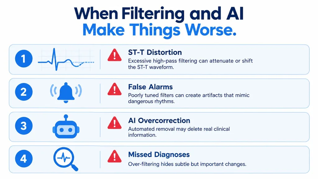
A filter can make a tracing look calmer without making it more accurate. Excessive high-pass filtering can attenuate or shift the ST-T waveform. Poorly designed filters can introduce phase distortion and alter QRS or repolarization measurements, which matters when clinicians assess ischemia or arrhythmia.
That creates a dangerous temptation. A clinician sees a smoother line and assumes the underlying signal is now trustworthy. In reality, the software may have suppressed information, reconstructed a waveform, or changed the appearance of clinically important morphology.
Quality control is useful, not infallible
An automated detector for nonphysiological artifacts and possible limb-lead misplacement reported 84.9% sensitivity, 100% specificity, and an ROC AUC of 0.924. Those results show that automated quality checks can identify unusable recordings, but they don't make the algorithm perfect. The reported detector evaluation also includes a 2025 preprint reporting 98.76% accuracy, 98.74% sensitivity, and 98.77% specificity for a deep-learning system classifying usable versus motion-corrupted resting ECGs. Those results are dataset-specific and shouldn't be treated as universal clinical performance.
Use a preserve, verify, repeat habit:
- Preserve: Keep the original tracing and don't overwrite the raw signal.
- Verify: Label suspected artifact and review the unprocessed signal when available.
- Document: Record the likely technical cause and the corrective action.
- Repeat: Obtain a new ECG after fixing the patient, electrode, cable, or environment.
Filtering can support interpretation, but it shouldn't conceal unresolved artifact when ischemia or a dangerous rhythm is under consideration. A visually impressive tracing isn't automatically a diagnostically reliable one.
Building Artifact Awareness Into Your Practice
Artifact recognition belongs in ACLS, PALS, BLS, telemetry, and routine bedside ECG workflows. In each setting, the same habits protect decision-making: correlate the monitor with the patient, identify the likely source, correct it, and document what changed.
Documentation helps the next clinician inherit context instead of a mystery strip. Record the suspected artifact, likely cause, corrective action, and final tracing quality. If the patient couldn't remain still or the signal remained limited, state that clearly rather than allowing a future reader to mistake technical uncertainty for a cardiac finding.
Make the skill repeatable
A short review routine can reinforce the habit:
- During ACLS: Confirm pulse, consciousness, and perfusion before treating an alarming waveform as a shockable rhythm.
- During PALS: Consider movement, crying, muscle activity, and electrode contact when a child's monitor changes abruptly.
- During BLS: Use the clinical assessment and organized resuscitation sequence rather than relying on a visually chaotic screen.
- During telemetry review: Compare suspicious segments with the patient's status and obtain a cleaner recording when the rhythm doesn't fit.
- During education: Practice identifying pattern, source, correction, and verification as one connected task.
Online CE and certification are valid, accessible options for healthcare professionals. A systematic review found that pure e-learning was no better or worse than face-to-face education for the primary outcomes assessed, while e-learning improved evidence-based knowledge and skills compared with no education. Blended learning also improved knowledge, skills, attitudes, and behavior compared with no learning. The systematic review of e-learning in evidence-based healthcare supports evaluating course quality and assessment rather than assuming classroom attendance is automatically superior.
A separate review of spaced digital education found a post-intervention knowledge advantage with a standardized mean difference of 0.32, with a 95% confidence interval of 0.13 to 0.51, and a knowledge-retention advantage with a standardized mean difference of 0.38, with a 95% confidence interval of 0.10 to 0.65. The review of spaced digital education supports repeated, structured learning over one concentrated session.
The broader industry is moving toward accreditation, meaningful assessment, curriculum quality, and practical outcomes. Hospitals, clinics, and professional organizations increasingly recognize accredited online CE and certification pathways, and peer-reviewed evidence doesn't support the outdated belief that only AHA or American Red Cross in-person classes are valid. Online and in-person formats can provide comparable educational outcomes, while online delivery adds scheduling flexibility and avoids travel or classroom release time. ProMed Certifications offers online ACLS, PALS, BLS, and Neonatal Resuscitation tracks that healthcare professionals can use to reinforce this framework through self-paced education.
ProMed Certifications offers online ACLS, PALS, BLS, and Neonatal Resuscitation courses designed for healthcare professionals who need flexible certification and review. Visit ProMed Certifications to connect your ECG artifact recognition practice with convenient continuing education and certification pathways.