

Treatment of Wide Complex Tachycardia: A Clinicial Guide
ECG interpretation is a critical component of bedside decision-making, and wide complex tachycardia is one of the rhythms that rewards calm thinking more than fast guessing. If you're at the monitor right now, the job is simple in theory and hard in practice, stabilize the patient, classify the rhythm, and choose the safest next move without turning a dangerous rhythm into a worse one.
That's why the treatment of wide complex tachycardia starts with a mindset shift. Online CE and certification formats can support that kind of learning just as well as in-person classes, because you can review rhythm strips, dosing, and decision pathways at your own pace, then revisit the parts that matter most at the bedside. That flexibility matters in real clinical life, and it's one reason accredited online education keeps gaining acceptance across hospitals, clinics, and professional teams.
Why Wide Complex Tachycardia Demands a Different Mindset
You're at the bedside, the monitor starts showing a broad QRS rhythm, and the room gets quiet for a second. One person is reaching for a medication, another is asking whether this could still be SVT, and the blood pressure tells you how much time you really have.
That is the right place to begin, because wide complex tachycardia is not just a faster narrow rhythm with a wider QRS. It is a separate emergency problem, and the first decision is whether the patient can tolerate a careful drug trial or needs immediate electrical treatment.
The bedside trade-off matters. A calm, structured approach helps you avoid giving the wrong drug to a rhythm that will not forgive it, while still moving quickly enough to protect perfusion. The ACLS tachycardia with a pulse algorithm is useful here because it keeps the first question anchored to stability, not to guesswork.
A practical analogy is an engine that starts knocking in bad weather. You do not keep testing every possible explanation before protecting the machine. You secure the patient first, then sort out the mechanism of the rhythm.
The emergency department sees this rhythm in a small slice of visits, but when it appears, it usually involves older patients and sicker presentations. One ED case series of 306 cases found WCT accounted for about 0.2% of all emergency department visits, with a mean age of 73.9 years, 66.0% male, 53.3% admitted, and 2.3% dying in the ED. That is why a structured bedside response matters after the first glance.
Once you see a wide rhythm, the next mistake is often trying to force every case into a single generic algorithm. Polymorphic VT and pre-excited AF have their own paths, and they should be treated as separate problems rather than variations of the same one.
Practical rule: If the patient looks unstable, do not let the rhythm label delay treatment. Perfusion status comes first.
The VT-First Default and How to Classify the Rhythm Quickly
The safest default is still the oldest one, assume VT until proven otherwise. The reason is simple. The 2019 ESC guideline says wide QRS tachycardias are typically VT about 80%, supraventricular with bundle-branch block aberrancy 15%, or antegrade accessory-pathway conduction 5%, and warns that treating VT as SVT can be harmful.
That means the first mental move is not “what drug do I like?” It's “which bucket is this rhythm most likely in, and how risky is my guess?”
The quick bedside split
Think in four questions:
- Is the patient stable? Blood pressure, mental status, chest pain, and pulmonary edema tell you whether you have time.
- Is the rhythm regular or irregular? Irregular wide rhythms raise the alarm for pre-excited atrial fibrillation.
- Is it monomorphic or polymorphic? Monomorphic rhythms tend to follow the VT pathway, while polymorphic rhythms open a different branch.
- Do I have a plausible SVT story? Prior history, clear triggers, and a narrow-complex baseline can matter, but they don't override danger.
In structural heart disease, the chance of VT is even higher than the general estimate, so a cautious stance is usually the safer one. If the rhythm is regular and monomorphic, you can think about pharmacology. If it's irregular or unstable, the room shifts toward shock therapy or urgent cardioversion. The “treat as VT first” habit pays off.
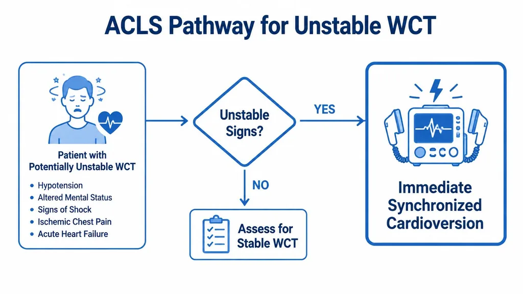
For a compact bedside reference, the ACLS tachycardia with a pulse algorithm is easy to keep close during review. If you're refreshing credentials, ACLS Certification is one online option clinicians use to work through rhythm logic on a flexible schedule.
Unstable Patients and Synchronized Cardioversion
If the patient is hypotensive, confused, having ischemic chest pain, or slipping into acute heart failure, the diagnosis can wait for later. The treatment can't.
The best bedside move is synchronized cardioversion for an unstable regular monomorphic WCT, because the problem isn't academic uncertainty, it's poor perfusion. Expert reviews emphasize that unstable patients should be cardioverted without delay, while stable regular WCT may only be approached with adenosine when SVT with aberrancy is strongly suspected source.
How to charge and shock
For monomorphic VT, the usual starting point is 100 joules synchronized, then escalate if the rhythm persists. The important detail is to stay in synchronized mode when the rhythm is organized enough to sense a QRS, because that reduces the risk of an R-on-T shock.
There's one major exception. If the rhythm is polymorphic and the patient is unstable or pulseless, synchronized cardioversion is the wrong tool. In that setting, go to defibrillation instead, because the rhythm is too chaotic for sync.
A practical way to frame the decision is this:
- Stable enough to think? Consider a cautious drug strategy if the rhythm fits.
- Not stable? Don't negotiate with the monitor, shock it.
For the bedside distinction between the two shock modalities, cardioversion and defibrillation is worth reviewing when the difference needs to be crystal clear.
Stable Monomorphic VT and the Procainamide Versus Amiodarone Choice
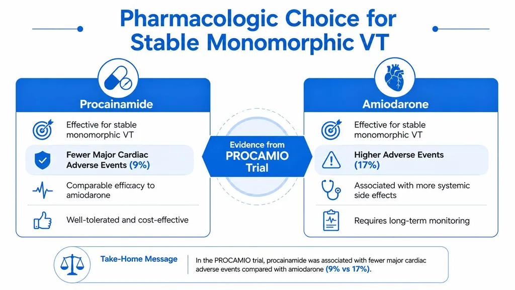
A stable, regular, monomorphic wide rhythm is the one place where a cautious drug trial makes sense. Here, procainamide has the strongest bedside case when blood pressure and perfusion are holding.
The key randomized benchmark is the PROCAMIO trial. In tolerated wide-QRS tachycardia, procainamide produced fewer major adverse cardiac events than amiodarone, with 9% vs 41% major events and more frequent tachycardia termination, 67% vs 38%. That's why many clinicians reach for procainamide first when the rhythm is regular, monomorphic, and the patient is stable.
Dosing that's usable at the bedside
A common protocol is 20 to 50 mg/min IV until one of four things happens, arrhythmia suppression, hypotension, QRS widening by more than 50%, or a total dose of 17 mg/kg. If the rhythm converts and the protocol continues, maintenance infusions are often 1 to 4 mg/min for about 6 hours in some settings.
That gives you clear stop rules, which matter more than enthusiasm for any one antiarrhythmic. If the pressure falls or the QRS widens significantly, stop the infusion and reassess.
Amiodarone still has a place, and major guidelines list it as an option for stable wide-complex tachycardia of unknown origin. A common loading dose is 150 mg IV, but the onset is slower, and the drug can be less attractive when you want a rapid termination attempt in a well-tolerated rhythm.
Bedside takeaway: Procainamide is the cleaner first choice for stable monomorphic VT when you can monitor continuously and stop fast if the patient worsens.
If you're reviewing antiarrhythmic selection for recertification, amiodarone guidance is a practical cross-reference for how clinicians often think about the drug in real practice.
When Adenosine and AV-Nodal Blockers Help or Hurt
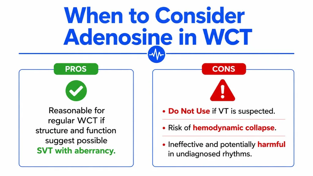
Adenosine feels tempting because it's fast, familiar, and often benign in the right rhythm. The problem is that wide complex tachycardia is not the right rhythm unless you have a strong reason to believe it's SVT with aberrancy.
That's the trap. Emergency guidance warns that AV-nodal blockers can precipitate collapse when the etiology of a wide-complex tachycardia is unestablished, and that irregular WCT consistent with pre-excited atrial fibrillation should be cardioverted rather than treated with routine nodal blockers source. In plain language, don't reach for the “small test dose” unless the worst-case scenario is one you can live with.
Where these drugs fit, and where they don't
Adenosine can be reasonable in a regular monomorphic WCT when the story strongly suggests SVT with aberrancy. Outside that narrow lane, it's a diagnostic and therapeutic gamble.
AV-nodal blockers are even more dangerous when pre-excitation is possible. Calcium channel blockers can worsen the situation in the wrong rhythm, which is why a drug like diltiazem deserves respect even when it's routinely used for other tachyarrhythmias. If you want a pharmacology refresher, Cardizem uses and side effects is a helpful background resource for the class.
The practical rule is short:
- Regular, monomorphic, and likely SVT with aberrancy? Adenosine may be reasonable.
- Irregular wide rhythm? Think pre-excited AF and cardiovert.
- Mechanism uncertain? Avoid AV-nodal blockers until the rhythm is clearer.
That's why bedside rhythm logic matters more than habit. A brief pause to classify the rhythm can prevent a fast deterioration.
Polymorphic VT, Torsades, and What Changes
Polymorphic wide-complex tachycardia is a different problem from monomorphic VT, and torsades de pointes sits at the center of that distinction. The mistake many clinicians make is trying to force every wide rhythm down the same treatment lane.
Torsades is treated with IV magnesium, and if the patient becomes unstable or degenerates, the right move is defibrillation rather than synchronized cardioversion. The rhythm often reflects a trigger problem, not just an electrical problem, so the bedside work has to include correction of QT prolongation, electrolyte abnormalities, or drug toxicity.
What to fix first
The cause matters as much as the shock. If the QT is prolonged, look hard at the medication list and any reversible contributors. If potassium or magnesium is low, repletion becomes part of the arrhythmia treatment, not a separate task.
This is also where the underlying substrate changes your thinking. Polymorphic VT in the setting of ischemia needs the ischemic problem addressed, while torsades from medication effect or electrolyte derangement needs that trigger removed. In both cases, treating the rhythm without treating the cause leaves the patient at risk for recurrence.
For physicians building CME around arrhythmia recognition, ProMed+ Physician CME is one online option that can fit into a busy schedule while covering rhythm management topics in a flexible format.
Clinical anchor: If the rhythm is polymorphic, don't default to the monomorphic VT playbook. Treat the trigger, and shock when instability demands it.
Diagnostic Workup and Disposition After the Rhythm Is Controlled
Once the rhythm is controlled, the focus shifts from rescue to sorting out risk. The next questions are why the patient developed WCT, whether there is an underlying substrate that makes recurrence more likely, and who needs monitoring, admission, or close follow-up.
Start with the bedside basics. Get a 12-lead ECG before and after conversion if it can be done safely, check a basic metabolic panel with potassium and magnesium, troponin, CBC, and a digoxin level when relevant. A focused echocardiogram helps when structural heart disease is on the table, because that changes how worried you should be about recurrence and how aggressively follow-up should be arranged.
A stable-looking rhythm can still hide a dangerous setup.
Who stays, who goes
Disposition should be deliberate, not improvised. The ED series of 306 WCT cases found 53.3% admitted and 2.3% died in the ED, which is a reminder that conversion does not automatically make the problem small.
A practical rule is straightforward:
- Admit or observe on telemetry if there is structural heart disease, recurrent VT, unexplained syncope, persistent symptoms, or abnormal labs.
- Refer to electrophysiology when the episode suggests re-entrant or scar-related arrhythmia, or when the diagnosis is still unclear.
- Consider discharge only after careful review when the episode was clearly stable, converted cleanly, and the evaluation does not suggest a dangerous substrate.
Medication review matters before the patient leaves. Check for QT-prolonging agents, AV-nodal blockers given by mistake, and drugs that can shift electrolytes. Clear return precautions should include chest pain, syncope, dyspnea, palpitations, and any recurrent rapid rhythm.
The rhythm branch also matters. Treat any WCT as VT first, shock the unstable patient immediately, use procainamide as the default for stable monomorphic VT, separate polymorphic rhythms into their own pathway, and avoid AV-nodal blockers when the mechanism is uncertain. For a focused review of polymorphic VT, PVT arrhythmia explained is a useful companion when you want that branch of the algorithm to feel cleaner at the bedside.
For clinicians who want a structured way to keep this material fresh, ProMed Certifications offers online ACLS and related courses you can work through on your schedule. The format is practical for reviewing tachycardia algorithms, dose choices, and rhythm strips before the next real case.