

Bundle Branch Block: Clear ECG Criteria for ACLS

ECG interpretation is a critical component of bedside care, and it's one of those skills that can change what happens in the next five minutes. If you're staring at a wide QRS on a monitor, you're not just looking at a tracing, you're making a judgment that affects whether the patient gets reassurance, more workup, pacing, or a trip to the cath lab. This guide will help you sort out bundle branch block with the same calm, stepwise approach used in ACLS, BLS, and PALS teaching, so you can separate a benign conduction delay from something that needs action.
A lot of clinicians learn ECGs in pieces, then get caught in the moment when the strip looks messy. That's where this topic matters most. A wide complex rhythm isn't automatically ventricular tachycardia, and a bundle branch block isn't automatically harmless. The skill is knowing when the pattern fits a stable conduction delay and when it's a warning sign that changes management.
A Wide QRS in Front of You
A code-team nurse glances at the monitor, sees a broad, slurred QRS, and has one job in that moment, avoid the wrong label. That's the bedside reality of bundle branch block, because a wide complex can pull you toward ventricular tachycardia when the patient may have a conduction delay. The difference matters, since a misread strip can delay the right response, while a new left bundle branch block in chest pain can hide a major ischemic problem.
The first thing to remember is that the ECG is a snapshot, not the diagnosis itself. The strip tells you what the electrical impulse looked like as it moved through the heart, but it doesn't tell you the whole story unless you pair it with symptoms, history, and the lead pattern. That's why the same tracing can be incidental in one patient and urgent in another.
Practical rule: Never call a wide QRS “just a BBB” until you've checked whether the patient is stable, symptomatic, or having chest pain, syncope, or heart failure symptoms.
That bedside habit is exactly why this topic shows up in certification exams and in real resuscitation rooms. The learner who understands the pattern can move faster, ask better questions, and avoid the two classic errors, overcalling ventricular tachycardia or undercalling a new conduction change. If you want a quick ECG refresher alongside this topic, the basic ACLS ECG interpretation guide is a useful companion.
The payoff is simple. Ten focused minutes on bundle branch block can improve how you read a strip at the bedside, how you document it, and how confidently you decide what happens next.
What Bundle Branch Block Actually Means
The heart's electrical system moves in a predictable path. The impulse starts in the SA node, reaches the AV node, then travels through the His bundle and splits into the right and left bundle branches before spreading through the Purkinje system. When one of these branches is slowed or blocked, the electrical signal has to take a longer alternative route.
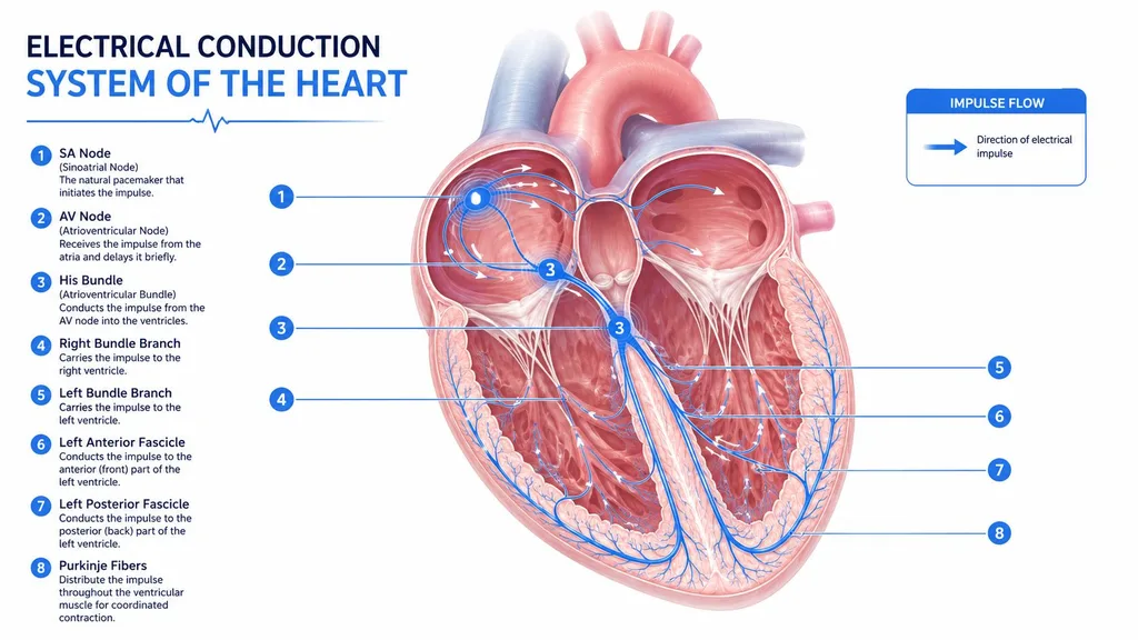
A bundle branch block is an electrical conduction delay in the heart, and it happens when one of those branches doesn't conduct normally. The signal still gets through, but it reaches part of the ventricle later, so the QRS looks wide. Mayo Clinic lists increasing age and underlying cardiovascular problems such as high blood pressure or heart disease as key risk factors, which fits what clinicians see at the bedside: this becomes more common as patients get older and as structural disease accumulates.
Right and left are not the same problem
A right bundle branch block means the right-sided conduction pathway is delayed. A left bundle branch block means the left-sided pathway is delayed. That difference matters because the left side is more often tied to structural heart disease and changes the way you think about chest pain, heart failure, and further testing.
Clinical anchor: BBB is a pattern on the ECG, not a disease by itself. The next question is always, “What caused it, and does it matter right now?”
That's the mindset that keeps the interpretation honest. A chronic pattern in an asymptomatic person may need little immediate action. A new pattern in a sick patient can be a clue that something much bigger is happening.
ECG Criteria for Right and Left Bundle Branch Block
The easiest way to read a bundle branch block is to compare the right and left patterns side by side. In practice, you start with the QRS width, then you look at V1, then you check the lateral leads. That sequence keeps you from getting lost in the noise.
The quick visual pattern
In RBBB, the right ventricle depolarizes late, so V1 often shows a terminal positive deflection and the lateral leads show a wide terminal S wave. In LBBB, the left ventricle depolarizes late, so V1 tends to go negative and the lateral leads show a broad, notched, often dominant R wave. The morphology is the clue, not just the width.
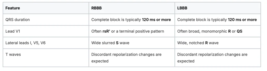
A useful anatomy reference for the chamber layout is the accurate cardiac muscle guide, because the bundle branch pattern makes more sense when you visualize which ventricle is being activated late.
What the bedside reader checks first
- Start with V1: It's often the fastest way to separate RBBB from LBBB.
- Then look at I, V5, and V6: The lateral leads usually confirm what V1 suggests.
- Check the QRS width: Wide alone isn't enough, but it sets the frame.
- Compare with a prior ECG: Chronic and new patterns don't carry the same weight.
If you're studying for credentialing and want a practical strip-reading workflow, the ProMed ECG interpretation walkthrough fits well here. For clinicians who want a certification option that can be completed online, ACLS Certification is one example of a course format that's designed to be quick and convenient, with the class coming directly to you and the card available in as quick as one hour or less.
Broad QRS width tells you there's a conduction problem. The lead pattern tells you which branch is delayed.
That's the difference between memorizing a rule and reading the strip.
Reading Real Strips Without Getting Lost
A healthy 28-year-old athlete can have an incomplete right bundle pattern and still be fine. In that setting, the strip may show a mild terminal r in V1 with a broader-looking S in the lateral leads, but the clinical picture stays calm because the patient is young, active, and asymptomatic. The key is that the ECG pattern must fit the person, not just the textbook.
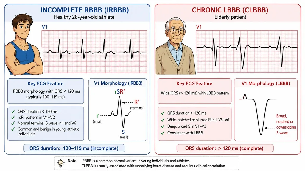
An elderly patient with a chronic left bundle pattern is a different read. The wide QRS, the broad lateral R waves, and the expected discordant repolarization changes are all more concerning if the tracing is new or the patient has dyspnea, fatigue, or chest discomfort. Chronicity matters because a stable pattern often reflects long-standing conduction disease rather than a new emergency.
A third strip can be trickier. A wide-complex tachycardia that looks like right bundle branch block may be SVT with aberrancy, which is why rhythm, rate, and clinical stability all matter together. A strip alone doesn't tell you whether the rhythm started above the ventricles and just conducted oddly, or whether the ventricles are driving the rhythm.
Before you interpret a real tracing, it helps to think through the context:
- Young athlete, no symptoms: incomplete RBBB can be incidental.
- Older patient, chronic disease: LBBB often deserves a closer look.
- Fast wide rhythm, unclear story: don't force a BBB label without checking the whole picture.
If your workflow includes bedside ECG supplies, it can also help to keep your equipment consistent.
Common Causes You Will Actually See
The causes that matter most are the ones you meet on rounds, in the ED, or during a rapid response call. Ischemic heart disease sits near the top of the list, especially when a new left bundle pattern appears in a patient with chest pain. Structural disease, chronic conduction system degeneration, pulmonary disease, congenital disease, and procedure-related injury can all show up as a bundle branch block pattern on the ECG.
The clinical buckets that stick
A patient with hypertensive heart disease may have long-standing conduction delay because the heart has remodeled under pressure load. Someone with cardiomyopathy may show a block because the conduction system sits inside diseased myocardium. A patient with cor pulmonale or pulmonary embolism can also develop conduction changes that make the strip look wide and abnormal.
Degenerative disease is another important category. Lenegre disease and Lev disease are classic examples of progressive conduction system disease, where the branches themselves deteriorate over time. Congenital heart disease matters too, including repaired tetralogy of Fallot, where baseline right-sided conduction delay is a familiar finding.
Procedures can trigger it as well. A right heart catheter or Swan-Ganz catheter can irritate the right bundle, so a new RBBB after line placement deserves attention. The same logic applies when a clinician sees an unexpected change after an intervention, because timing often points to the cause.
Bedside pearl: When a bundle pattern appears right after a catheter passes through the right heart, think mechanical irritation first, not coincidence.
For learners in hybrid practice settings, this is also where remote work and cardiology review overlap.
The main lesson is simple. Don't memorize causes as a flat list. Group them by the kind of patient in front of you, and the strip becomes much easier to interpret.
Why Bundle Branch Block Matters in ACLS, BLS, and PALS
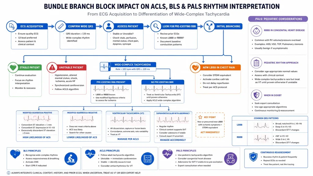
In ACLS, a pre-existing bundle branch block can make a wide rhythm harder to sort from ventricular tachycardia. That's why history matters during rhythm interpretation. If the patient has a known baseline BBB and now shows a similar wide pattern, you think differently than you would with a new wide-complex rhythm in an unstable patient.
The decision points that change care
A new left bundle branch block in a patient with chest pain raises concern for acute ischemia and should not be brushed off as a benign ECG variant. In contrast, a stable, longstanding block in an otherwise well patient may be an incidental finding. That distinction is the heart of bedside decision-making.
In heart failure, the stakes can be higher. In the CHARM Programme, cardiovascular death or heart failure hospitalization occurred in 42.6% of patients with BBB versus 18.6% in those with a normal ECG, and the authors describe BBB as an independent predictor of worse outcomes. That doesn't mean every block is dangerous, but it does mean the ECG can carry prognostic information that shapes follow-up and intensity of care.
PALS adds another layer. In children, BBB patterns often point you toward congenital or postsurgical heart disease rather than the same degenerative causes you expect in adults. That makes the context different, even when the tracing looks familiar.
If bradycardia management is part of your study plan, the ACLS bradycardia algorithm reference is a practical place to connect rhythm interpretation with treatment decisions. For clinicians who want a fully online pediatric option, PALS Certification is offered as a 100% online format that lets you finish the review, take the exam, and receive the card in as quick as one hour or less.
Certification takeaway: BBB matters most when it changes what you do next, not when it just changes how the strip looks.
That's why it belongs in ACLS, BLS, and PALS thinking. You're not only naming a pattern, you're deciding whether the pattern is part of the emergency.
Mimics That Fool Even Experienced Clinicians
Not every wide QRS is a bundle branch block. That's the trap. A broad complex can come from hyperkalemia, sodium-channel blocker toxicity, ventricular pacing, idiopathic ventricular rhythms, or pre-excitation such as WPW, and each one carries a different clinical response.
The short differential check
Start with the context, because the story often gives you the answer before the ECG does.
- Renal failure or severe metabolic illness: think hyperkalemia first.
- Known antiarrhythmic or toxic exposure: widen your differential to sodium-channel blockade.
- Pacemaker on the chest x-ray or visible spikes: consider a paced rhythm.
- Young patient with very fast wide tachycardia: don't assume BBB, look for WPW or ventricular origin.
- Syncope, instability, or ischemic symptoms: keep ventricular rhythms high on the list.
Hyperkalemia often gives you more than a wide QRS. You may also see peaked T waves and progressive conduction slowing. A paced rhythm has a different visual signature, because the pacemaker spike changes the rhythm logic before the QRS even begins. WPW can be especially misleading when a fast rhythm makes the complex appear broad and odd.
If the ECG and the story don't agree, pause and widen the differential before you call it a bundle branch block.
That caution saves time and keeps you from anchoring on the first label that fits part of the tracing. In emergency care, a wide QRS is a clue, not the verdict.
Follow-Up, Management, and Study Next Steps
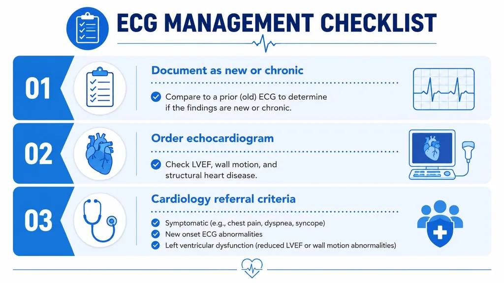
The first management step is to decide whether the pattern is new or chronic. Compare the tracing with an old ECG if one exists, and document the difference clearly. That single comparison often tells you whether you're dealing with a stable conduction pattern or a fresh change that deserves more attention.
The practical checklist
- Document the timing. Note whether the BBB is new, old, intermittent, or previously known.
- Match the strip to symptoms. Chest pain, syncope, dyspnea, and heart failure signs all raise the stakes.
- Look for structural disease. An echocardiogram is often the next logical test when the pattern is new, unexplained, or paired with concerning symptoms.
- Refer when needed. Cardiology involvement makes sense when the BBB is symptomatic, new, associated with left ventricular dysfunction, or part of a broader conduction problem.
- Think ahead about pacing or resynchronization. Some patients with conduction disease eventually need device-based therapy, especially when the block reflects more advanced electrical disease.
The follow-up question is not just “what is it?” It's “what does it mean for this patient today?” That's the same question clinicians ask during certification review, because exam writers love cases where the tracing looks simple but the clinical context changes everything.
For learners balancing work and study, online CE and certification formats have become a practical part of modern healthcare education. High-quality online courses can be completed on a clinician's schedule, and many organizations now treat accredited online options as a normal part of ongoing training rather than a second-tier substitute. ProMed Certifications offers an online pathway for ACLS, BLS, and PALS that fits busy schedules, which is useful when you're reviewing ECGs and need the flexibility to study between shifts.
The core idea is steady and simple. Know the pattern, check the context, document the chronology, and escalate when the patient or the ECG tells you the block is more than incidental.
If you want to strengthen your ECG rhythm recognition and keep your certification study flexible, visit ProMed Certifications to review online ACLS, BLS, and PALS options that fit a clinician's schedule. It's a practical way to keep your skills sharp while you build confidence with bundle branch block interpretation at the bedside.